
In vivo metastatic prostate tumor model development and follow-up progression using in vivo bioluminescence imaging.
Authors
Vincent Faugeroux, Nicolas Hoffmann, Sarah Belderbos, Maëva Albanese, Kenny Herry, Nicolas Ancellin, Caroline Mignard, Marc Hillairet De Boisferon
Oncodesign Services – 20 rue Jean Mazen, 21000, Dijon, France
Abstract
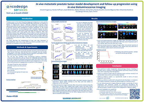
Prostate cancer is the most frequently diagnosed malignancy in men and the second leading cause of cancer-related death after lung cancer. Its incidence is highest in high-income regions. Although widespread screening allows approximately 80% of cases to be detected while still organ-confined, 15–20% of patients present with locoregional or distant metastases. In the United States, the proportion of men diagnosed with metastatic disease has been rising in recent years, particularly among younger patients. Clinical outcome strongly correlates with tumor grade and stage at diagnosis; men with metastatic disease have significantly reduced overall survival compared with those with localized tumors. The primary metastatic sites are lymph nodes and bone, followed by liver and lung. However, few preclinical models effectively recapitulate these advanced stages, creating a critical gap for evaluating novel therapeutics.
Here, we describe the establishment of liver and lung metastases in immunodeficient male mice using 22Rv1-Luc-mCherry prostate cancer cells delivered via intratibial or intra-arterial injection. Mice were monitored three times per week for clinical condition and body weight. Tumor progression was assessed by in vivo bioluminescence imaging once per week for five weeks. At euthanasia, major organs were collected for ex vivo imaging to confirm metastatic localization.
Following intratibial inoculation, bioluminescence increased initially at the injection site during the first two weeks, then predominantly in the liver and lungs from days 15 to 42. These findings were confirmed by ex vivo imaging. After intra-arterial inoculation, early bioluminescence was detected primarily in bone through day 28, followed by a marked increase in the liver between days 28 and 35. Ex vivo imaging verified metastatic dissemination to the liver, limb bones, spinal column, and seminal vesicles.
In summary, we report a reproducible metastatic prostate cancer model that mimics key features of late-stage disease, providing a valuable platform for preclinical evaluation of new therapeutic compounds.