
What bioluminescence imaging reveals when study design comes first.
Bioluminescence imaging (BLI) is a mainstay of preclinical research, valued for its sensitivity and ability to support longitudinal studies in living systems. It offers a powerful way to visualise biological processes as they unfold. By allowing biological activity to be tracked repeatedly in the same living system, bioluminescence imaging reveals when, where, and how biological processes evolve over time – information that is critical for interpreting preclinical efficacy and mechanism.
In practice, successful BLI is rarely plug-and-play. Extracting meaningful insight depends on how thoughtfully experimental design and substrate selection are matched to the underlying biology. Rather than a single technical decision, substrate choice often becomes part of the discovery process itself.
Enzyme-substrate characteristics shape what you can see.
Not all luciferase–substrate systems behave the same way in vivo. Emission wavelength, tissue penetration, and signal intensity all influence whether biologically relevant activity is detectable.
Classical firefly luciferase remains a highly accessible and cost-effective option, and in many contexts performs exceptionally well. However, as studies move toward lower expression levels, deeper tissues, or more subtle biological effects, signal attenuation can become a limiting factor. In the study example below, alternative substrates with red-shifted emission profiles were explored to improve detectability. While these systems required additional optimization and came at a higher cost, they enabled clearer signal discrimination that would likely have been missed using a standard approach.
Importantly, the biology had not changed, only the ability to observe it had improved.
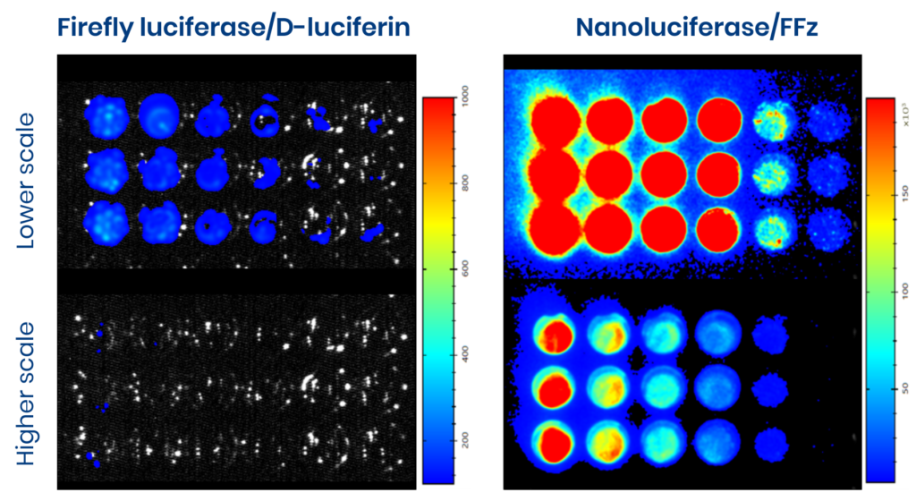
Above: Bioluminescence imaging of a cell line transduced for a vector encoding for firefly luciferase (left) or nanoluciferase (right) at two different scales. In the same conditions, higher bioluminescent signal is detected for cells expressing for nanoluciferase after addition of FFz compared to those expressing firefly luciferase (substrate D-luciferin).
Balancing sensitivity, cost, and scientific need.
Higher-performance substrates are not universally better. They typically require greater investment and more careful experimental control. The decision to use them should therefore be driven by scientific necessity rather than default preference.
If a conventional system reliably answers the research question, simplicity has clear advantages. But where sensitivity is limiting, investing in a more advanced substrate can prevent false negatives and avoid premature conclusions about biological inactivity.
Starting with kinetics when building a study.
Early-stage kinetic evaluation is one of the most informative steps in BLI study design. Understanding how rapidly a substrate distributes, how stable the signal remains over time, and when a reproducible plateau is reached allows us to define acquisition windows with confidence.
In applied studies, these early experiments frequently reveal behaviour that would not be predicted from datasheets alone. For example, in one recent longitudinal in vivo study at Oncodesign Services, multiple substrate concentrations were evaluated during initial optimization. While lower doses appeared sufficient at first glance, kinetic profiling showed unstable signal behaviour and poor reproducibility. Higher concentrations produced more consistent plateau phases but also revealed increased inter-animal variability that only became apparent when individual curves were examined.
This type of insight (missed if optimization is skipped or averaged too early) often determines whether downstream data can be interpreted with confidence.
Aligning study design with technical capability to drive discovery.
Robust BLI studies benefit from strategies that reduce confounding signal and improve interpretability. This may include separating regions of interest during acquisition, controlling background emission, or designing workflows that allow multiple imaging perspectives within a single experiment.
Together, these choices elevate bioluminescence imaging from a confirmatory tool to a discovery-enabling one.
High-quality bioluminescence data does not come from technology alone. It emerges from the alignment of biological objectives, experimental design, and substrate performance. When substrate selection is treated as part of the discovery process rather than an afterthought, researchers gain clearer insight, reduce uncertainty, and make more informed decisions throughout preclinical development.
Key takeaways:
- Bioluminescence imaging is not a one-size-fits-all technique. Study design and substrate choice must be tailored to the biology and sensitivity requirements of each program.
- Early kinetic evaluation is critical. Dose-ranging and time-course studies help identify stable acquisition windows and prevent variability from undermining downstream data.
- Substrate performance directly affects detectability. Emission wavelength, signal stability, and tissue penetration determine whether biologically relevant signals can be confidently observed.
- Classical systems remain valuable but have limits. Firefly luciferase is cost-effective and robust for many applications but may miss low-level or challenging signals.
Find out more about our bioluminescent and in vivo imaging services here.