
Preclinical orthotopic tumors model can better recapitulate the tumor microenvironment (TME)
An orthotopic model is a type of preclinical cancer model where tumor cells are implanted into the original organ site, allowing researchers to study growth in a more physiologically relevant environment.
While many CDX and PDX tumors models grow subcutaneously in mice, sometimes a tumor model requires a specific kind of tumor microenvironment that can only be well recapitulated in the stroma of the primary organ. Orthotopic models improve physiological relevance by growing the tumor in the correct microenvironment to improve modeling local and distant metastatic invasion, drug exposure and local signaling pathway.
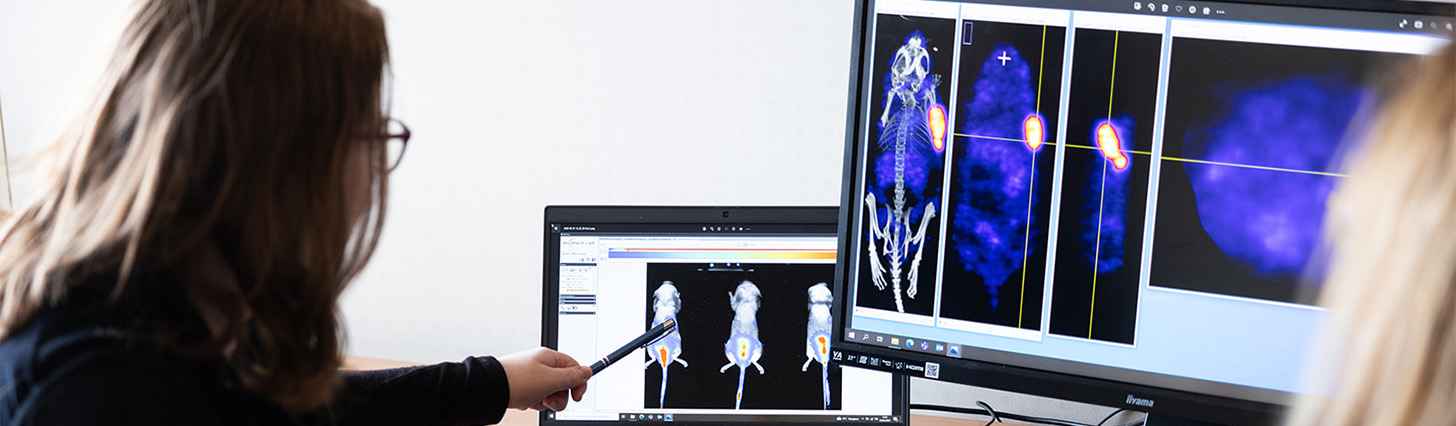
Oncodesign Services has surgery and imaging (including MRI, PET, PET/MRI, SPECT and optical imaging) capabilities which are suitable for orthotopic models. MRI and bioluminescence are typically used to size tumors for randomization and for monitoring tumor burden.
Exemple of available orthotopic models:
| Name | Organ | Histological Type | Host rodent species |
| SYNGENEIC | |||
| 4T1 | Breast | Carcinoma transitional | Balb/c or NSG Mouse |
| AY27 | Bladder | Carcinoma | F344 Fischer Rat |
| C6 | Brain | Glioma | Nude Rat |
| CT26 | Colon | Carcinoma | Balb/c Mouse |
| EMT6 | Breast | Mammary Carcinoma | Balb/c Mouse |
| GS-9L | Brain | Gliosarcoma | F344 Fischer Rat |
| GV1A1 | Brain | Glioma | BDIX Rat |
| Hepa1-6 | Liver | Hepatocarcinoma | C57BI/6 Mouse |
| MBT-2 | Bladder | Carcinoma | C3H Mouse |
| NBT-II | Bladder | Tumor | Wistar Rat |
| NCTC 2472 | Bone | Sarcoma | C3H Mouse |
| R3327H | Prostate | Adenocarcinoma | Copenhagen Rat |
| Renca | Kidney | Carcinoma | Balb/c Mouse |
| XENOGRAFT | |||
| LS 174T | Colon | Colorectal Adenocarcinoma | Nude Rat or Mouse |
| MDA-MB-175 | Breast | Adenocarcinoma | SCID Mouse |
| NCI-H146 | Lung | carcinoma-SCLC | Nude Rat |
| NCI-H209 | Lung | Carcinoma SCLC | Nude or SDRG Rat |
| NCI-H460 | Lung | Carcinoma-NSCLC | Nude Mouse |
| NIH:OVCAR-3 | Ovary | Adenocarcinoma | Nude Rat or Balb/C Nude Mouse |
| PANC-1 | Pancreas | Carcinoma | Nude Rat |
| PC-3 | Prostate | Adenocarcinoma | Nude Rat |
| PC-3-MM2 | Prostate | Adenocarcinoma | Nude Rat |
| RT-4 (CVE-4) | Bladder | Transitional papilloma | Nude Rat |
| SW-620 | Colon | Adenocarcinoma | SCID Mouse |
| U-87 MG | Brain | Glioblastoma | Swiss or NMRI Nude Mouse or Nude Rat |
Case Studies with Orthotopic Models
Model example #1
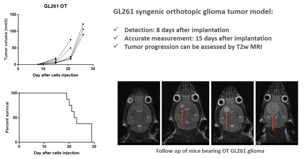
To assess the efficacy of immunotherapies in a physiologically relevant setting, Oncodesign Services developed an orthotopic glioblastoma model using the murine GL261 cell line implanted directly into the brain of mice. This model preserves the integrity of the tumor microenvironment and immune system, making it highly suitable for evaluating immune-modulating agents. In this study, tumors were detected 8 days after implantation using non-invasive techniques, allowing for precise tracking of intracranial tumor growth. The tumors were quantifiable with adequate animal health metrics for at least 15 days, with survival dropping after day 20.
Model example #2
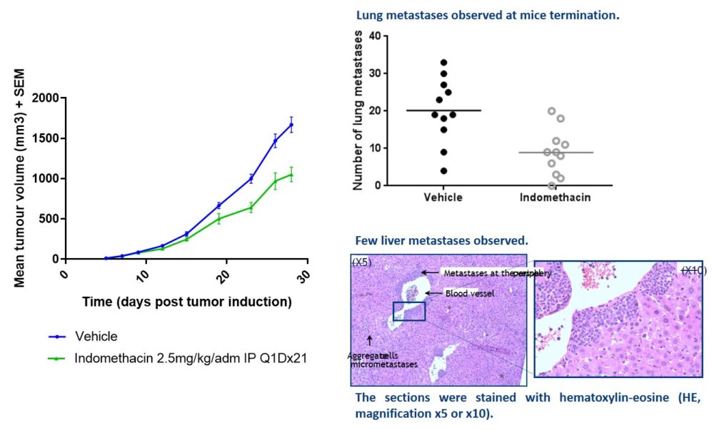
In this study, the 4T1 mouse mammary carcinoma model was orthotopically injected into the mammary fat pad (MFP) of immunocompetent BALB/c mice to replicate the tumor’s natural microenvironment and metastatic behavior. This syngeneic model is known for its aggressive growth and spontaneous metastasis, particularly to the lungs. Tumor size was monitored using caliper and total tumor burden was counted after termination.