


Preclinical models to support therapeutic assessment for liver injury, fibrosis and NASH/MASH.
Oncodesign Services provides preclinical liver models for investigating acute injury, liver fibrosis, and metabolic liver disease. Our scientists can help you select the model, disease stage, and readouts that best reflect your molecule’s mechanism of action, enabling translatable assessment of therapeutic effects on metabolic dysfunction, inflammation, fibrosis, and/or liver injury.
Feasibility and study design for liver-related disease programs
Acute and chronic inflammatory diseases of the liver can be triggered by several causes such as exposure to toxic chemicals, diet, or metabolic or immune dysregulation. Rodent models of liver inflammation are helpful in understanding the triggers and mechanisms of inflammatory liver diseases, and the therapeutic response expected in human patients.
Confirm feasibility before study initiation:
To support confident model selection, we also offer histology samples from established research models that can be accessed ahead of full study commitment to confirm feasibility. Immunohistochemistry (IHC) or FISH analyses may be performed to assess target expression and tissue localization, helping confirm biological relevance before program initiation. We currently offer the following samples for liver disease studies:
- NASH model of high fat diet + CCl4
- NASH model of streptozotocin and high fat diet
- CCl4 liver fibrosis
Typical readouts for liver inflammation studies:
- Histology:
- Steatosis
- Fibrosis
- Ballooning degeneration of hepatocytes
- Necrosis
- NASH score
- Serum markers of liver injury: LDH, ALT, AST
- Inflammation markers (e.g. cytokines)
- Immune cell infiltrates (flow cytometry, IHC)
- Apoptosis markers
- Gene expression in liver by qPCR/dPCR
- Biomarker / drug monitoring
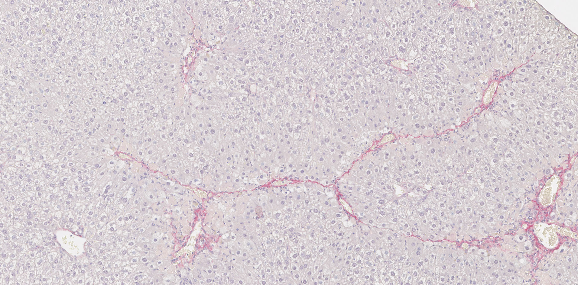
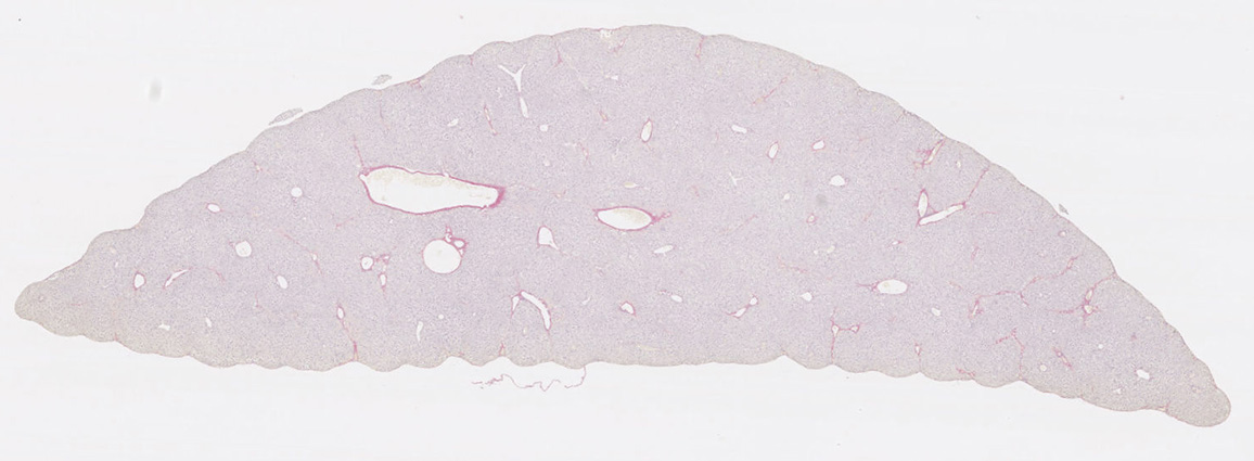
Histology slide of the liver fibrosis CCl4 model on D42 (Sirius Red stain). Here we can see collagen deposition and constricting structures.
Established liver inflammation models available for preclinical studies:
-
Liver Fibrosis Models
CCl4 model is a well-established liver fibrosis model for evaluating compounds that act directly on fibrotic and inflammatory processes. Unlike metabolic liver disease models, the CCl4 model does not include a dietary or metabolic component. It is therefore particularly relevant when the primary research question concerns the prevention, reduction, or resolution of liver fibrosis.
In the CCl4 mouse model, repeated exposure to carbon tetrachloride produces hepatocellular injury and inflammation, followed by progressive fibrotic tissue deposition. Studies can be designed over several weeks to investigate therapeutic activity at an appropriate stage of fibrosis development. This model can be especially useful for distinguishing a compound’s direct antifibrotic effects from activity related to steatosis or metabolic dysfunction.
Histological assessment is the gold standard in evaluating efficacy in a CCl4 liver fibrosis model. Sirius Red staining (or other collagen-specific stains), supported by digital image analysis and review by a pathologist, can be used to quantify collagen deposition and the distribution of fibrotic regions throughout the liver. Additional immunohistochemistry can localize relevant immune-cell populations, pharmacological targets, or markers of tissue remodeling.
Serum measurements such as AST, ALT, and GGT can provide complementary evidence of hepatocellular distress. Because these parameters may paradoxically decrease as hepatocytes progressively die, longitudinal or appropriately timed measurements are generally more informative than an isolated endpoint. Cytokine analysis, gene-expression studies, and immune profiling of circulating or splenic cells can also be incorporated according to the compound’s mechanism of action.
Available models:
- CCl4 fibrosis
-
NASH / MASH Models
Metabolic dysfunction-associated steatohepatitis, or MASH, is the current term for the disease previously known as non-alcoholic steatohepatitis, or NASH. Both terms remain widely used in preclinical research. A suitable NASH model or MASH model should reproduce the combination of metabolic dysfunction, hepatic fat accumulation, inflammation, and fibrosis that characterizes progressive human disease. An additional feature is the capacity of the models to reproduce the terminal evolution towards hepatocarcinoma development, especially in males.
We offer different NASH / MASH mouse model approaches to accommodate distinct mechanisms of action and development objectives. A shorter-duration model combines a high-fat, high-fructose diet with CCl4. The dietary component induces metabolic stress and steatosis, while CCl4 promotes the development of fibrosis. This model enables metabolic and fibrotic effects to be investigated within the same study and may be suitable for compounds targeting strictly meyabolic mechanisms or both components of the disease.
A second approach combines streptozotocin-induced type I diabetes with a high-fat high-fructose diet. This progressive MASH mouse model develops through clinically relevant stages that can include steatosis, inflammation, fibrosis, cirrhosis, and, at later timepoints, hepatocellular carcinoma in males. As its liver pathology develops primarily through metabolic drivers rather than a direct profibrotic agent, the model can provide a useful system for investigating disease progression and therapeutic efficacy at different stages, particularly for compounds targeting metabolic dysregulation. Its longer duration should be considered when selecting it for a particular program.
Histology is the primary method for assessing steatosis and fibrosis in NASH / MASH models. H&E and Sirius Red staining, digital quantification, and pathological review can provide a detailed assessment of liver morphology, lipid accumulation, inflammation, and collagen deposition. Depending on the stage and objectives of the study, later pathological changes and tumor development may also be assessed.
Metabolic measurements can help establish whether treatment is addressing the underlying dysfunction rather than fibrosis alone. These may include blood glucose, glucose-tolerance testing, circulating triglycerides, and other relevant biochemical parameters. AST, ALT, and GGT can support the assessment of hepatocellular injury, while cytokine profiling, gene-expression analysis, immunohistochemistry, and immune-cell profiling can provide further mechanistic insight. Readouts are selected according to whether the candidate is expected to act predominantly on metabolism, inflammation, fibrosis, or multiple disease processes.
Available NASH / MASH models:
- STZ+HFD NASH
- CCl4+HFD NASH
- Long term NASH-HCC
-
Acute Liver Injury Models
Acute liver injury models address a different biological and therapeutic question from chronic liver fibrosis and MASH models. These reproduce rapidly developing hepatocellular injury and inflammatory responses, enabling candidate therapies to be assessed for their ability to protect liver tissue or limit the progression of acute damage.
An acetaminophen-induced model can be used to investigate acute liver injury associated with drug toxicity. Acetaminophen, also known as paracetamol, is a clinically relevant cause of acute liver failure when taken in overdose. The model can support the evaluation of hepatoprotective candidates and treatments intended to reduce cellular injury following an acute toxic insult.
A complementary model uses Fas ligand, or CD178, or FasL/CD95L, to activate death-receptor signaling and induce acute hepatic injury. This provides an alternative means of investigating compounds that may regulate apoptosis, inflammatory signaling, or other mechanisms involved in acute liver damage.
Study endpoints can include histopathological assessment of the extent and distribution of liver injury, together with serum markers of hepatocellular damage. Apoptosis and necrosis may be evaluated using suitable tissue-based methods, while inflammatory responses can be characterized through cytokine analysis, including markers associated with TNF-family signaling. The final study design and readout panel are adapted to the candidate’s proposed mechanism of action and the aspect of acute liver injury being investigated.
Available acute liver injury models:
- FasL-induced acute liver injury model
- Acetaminophen-induced acute liver injury model
Experimental example: Using an ALK5 inhibitor as a reference compound in CCl4 liver fibrosis models
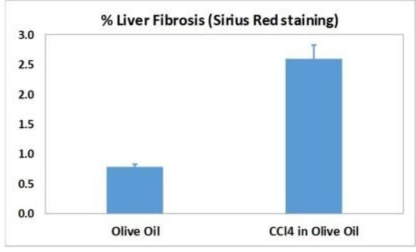 The CCl4 model of liver fibrosis is a classic model widely used in industry. Mice are given repeated doses of CCl4. Liver fibrosis typically appears at 6 weeks.
This model is useful for therapies aiming to modulate collagen depositions, expression of metalloproteases, and fibrotic processes. An ALK5 (TGFb receptor I) inhibitor, targeting a well-known anti-fibrotic mechanism, decreases collagen induction and is used as a reference compound.
In this bar graph (right), we can see CCl4-induced liver fibrosis biomarker results comparing the concentration of collagen fibers.
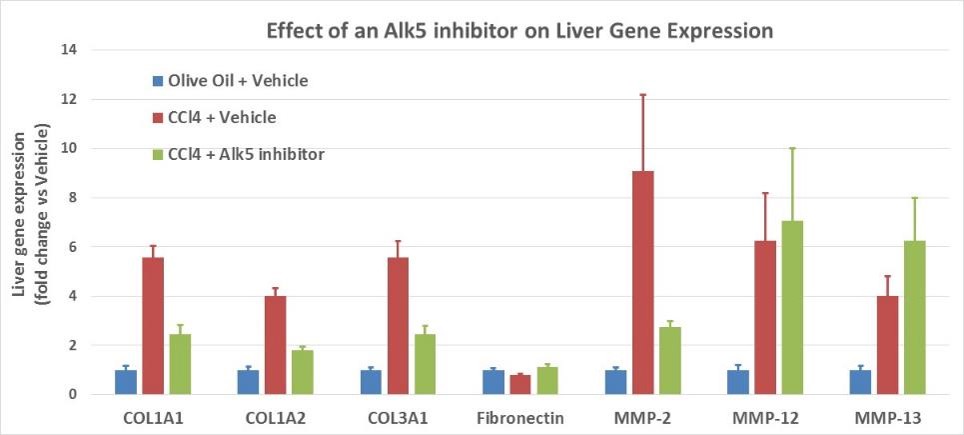
Here we see the comparative gene expression ALK5 inhibitor response in the CCl4 liver fibrosis model.
Learn more about partnering with Oncodesign Services:
Oncodesign Services combines translational inflammation experience with a flexible approach to preclinical research, supporting both standard and customized study designs. Alongside an extensive portfolio of acute and chronic inflammatory disease models, we offer bespoke model development to address specific scientific questions and emerging therapeutic approaches.
Our scientific team is available to provide guidance from study planning through data interpretation, with comprehensive readouts including histology, clinical scoring, biomarkers, and functional endpoints. Histology samples are also available to support feasibility assessments and model selection before study initiation.
Contact our team to discuss your research objectives, explore the most appropriate models and endpoints, or request a quotation.
Oncodesign Services (ODS) has been a trusted preclinical partner for several years. The team consistently demonstrates scientific excellence and reliability, tailoring preclinical protocols to our exact needs.
Their ability to rapidly action and deliver projects has made collaboration in oncology research efficient and straightforward. Clear, responsive communication is crucial to us, and the strong working relationship with our client manager makes coordination very easy.
We appreciate the professionalism, confidence, and flexibility ODS brings to every project.
Oncology Biopharma Organization (Europe)

Frequently asked questions about our liver disease models:
Which therapeutic modalities can you evaluate in liver disease models?
We support preclinical evaluation of a wide range of therapeutic approaches, including:
- Small molecules
- Monoclonal antibodies
- Peptides
- RNA therapeutics
- Cell and gene therapies
- Combination therapies
- Novel therapeutic platforms
Study designs are customized to align with your therapeutic strategy and development stage.
What endpoints do you measure in liver disease studies?
We offer comprehensive efficacy and mechanistic endpoints, including:
- Liver histopathology
- Fibrosis quantification
- Steatosis scoring
- Inflammation assessment
- Serum liver enzymes (ALT, AST)
- Lipid profiling
- Cytokine and chemokine analysis
- Gene and protein expression
- Immunohistochemistry
- Flow cytometry
- Digital pathology
- Multiplex biomarker analysis
Endpoints can be selected to match your target biology and regulatory objectives.
Can liver disease studies be customized?
Yes. Every study can be tailored to your program, including:
- Disease induction protocol
- Treatment initiation (preventive or therapeutic)
- Dosing schedule
- Route of administration
- Study duration
- Biomarker panel
- Tissue collection strategy
- Specialized mechanistic analyses
Do you provide study design support?
Yes. Our scientific team collaborates with sponsors to optimize study design, including:
- Model selection
- Treatment schedule
- Randomization and blinding strategy
- Endpoint selection
- Biomarker recommendations
-
References
(1) Acute liver injury models in mice
FasL:
Shao R, Yang Y, Fan K, Wu X, Jiang R, Tang L, Li L, Shen Y, Liu G, Zhang L. REV-ERBα Agonist GSK4112 attenuates Fas-induced Acute Hepatic Damage in Mice. Int J Med Sci. 2021 Oct 25;18(16):3831-3838. doi: 10.7150/ijms.52011. PMID: 34790059; PMCID: PMC8579287. https://www.medsci.org/v18p3831.htm
Ogasawara J, Watanabe-Fukunaga R, Adachi M, Matsuzawa A, Kasugai T, Kitamura Y, Itoh N, Suda T, Nagata S. Lethal effect of the anti-Fas antibody in mice. Nature. 1993 Aug 26;364(6440):806-9. doi: 10.1038/364806a0. Erratum in: Nature 1993 Oct 7;365(6446):568. PMID: 7689176 https://www.nature.com/articles/364806a0
Lesnikov V, Gorden N, Fausto N, Spaulding E, Campbell J, Shulman H, Fleming RE, Deeg HJ. Transferrin fails to provide protection against Fas-induced hepatic injury in mice with deletion of functional transferrin-receptor type 2. Apoptosis. 2008 Aug;13(8):1005-12. doi: 10.1007/s10495-008-0233-6. PMID: 18561026; PMCID: PMC2574612. https://link.springer.com/article/10.1007/s10495-008-0233-6
Ohtaki Y, Yamaguchi K, Yu Z, Kumamoto H, Shimauchi H, Iwakura Y, Sugawara S, Endo Y. Hepatic platelet accumulation in Fas-mediated hepatitis in mice. Int Immunopharmacol. 2009 Aug;9(9):1071-8. doi: 10.1016/j.intimp.2009.04.016. Epub 2009 May 9. PMID: 19439197. https://www.sciencedirect.com/science/article/pii/S1567576909001659?via%3Dihub
Donthamsetty S, Mars WM, Orr A, Wu C, Michalopoulos GK. Protection against Fas-induced fulminant hepatic failure in liver specific integrin linked kinase knockout mice. Comp Hepatol. 2011 Nov 21;10:11. doi: 10.1186/1476-5926-10-11. PMID: 22104495; PMCID: PMC3228663. https://www.ncbi.nlm.nih.gov/pmc/articles/PMC3228663/
Acetaminophen:
- Mechanism:
Ni HM, McGill MR, Chao X, Du K, Williams JA, Xie Y, Jaeschke H, Ding WX. Removal of acetaminophen protein adducts by autophagy protects against acetaminophen-induced liver injury in mice. J Hepatol. 2016 Aug;65(2):354-62. doi: 10.1016/j.jhep.2016.04.025. Epub 2016 May 2. PMID: 27151180; PMCID: PMC4955750. https://www.journal-of-hepatology.eu/article/S0168-8278(16)30169-6/fulltext
Groeneveld D, Cline-Fedewa H, Baker KS, Williams KJ, Roth RA, Mittermeier K, Lisman T, Palumbo JS, Luyendyk JP. Von Willebrand factor delays liver repair after acetaminophen-induced acute liver injury in mice. J Hepatol. 2020 Jan;72(1):146-155. doi: 10.1016/j.jhep.2019.09.030. Epub 2019 Oct 10. PMID: 31606553; PMCID: PMC6941657. https://www.journal-of-hepatology.eu/article/S0168-8278(19)30600-2/fulltext
- Review:
Mossanen JC, Tacke F. Acetaminophen-induced acute liver injury in mice. Lab Anim. 2015 Apr;49(1 Suppl):30-6. doi: 10.1177/0023677215570992. PMID: 25835736. https://journals.sagepub.com/doi/10.1177/0023677215570992?url_ver=Z39.88-2003&rfr_id=ori:rid:crossref.org&rfr_dat=cr_pub%20%200pubmed
osner I, Romero-Ferret C, Mottot G. Letter: Treatment of acute paracetamol poisoning. Lancet. 1973 Dec 1;2(7840):1273-4. doi: 10.1016/s0140-6736(73)91024-6. PMID: 4128608 https://www.thelancet.com/journals/lancet/article/PIIS0140-6736(73)91024-6/fulltext
- Testing compounds :
Jaeschke H, Akakpo JY, Umbaugh DS, Ramachandran A. Novel Therapeutic Approaches Against Acetaminophen-induced Liver Injury and Acute Liver Failure. Toxicol Sci. 2020 Apr 1;174(2):159-167. doi: 10.1093/toxsci/kfaa002. PMID: 31926003; PMCID: PMC7098369. https://academic.oup.com/toxsci/article/174/2/159/5700748?login=false
Niu B, Lei X, Xu Q, Ju Y, Xu D, Mao L, Li J, Zheng Y, Sun N, Zhang X, Mao Y, Li X. Protecting mitochondria via inhibiting VDAC1 oligomerization alleviates ferroptosis in acetaminophen-induced acute liver injury. Cell Biol Toxicol. 2022 Jun;38(3):505-530. doi: 10.1007/s10565-021-09624-x. Epub 2021 Aug 17. PMID: 34401974. https://link.springer.com/article/10.1007/s10565-021-09624-x
- Role of microbiota:
Schneider KM, Elfers C, Ghallab A, Schneider CV, Galvez EJC, Mohs A, Gui W, Candels LS, Wirtz TH, Zuehlke S, Spiteller M, Myllys M, Roulet A, Ouzerdine A, Lelouvier B, Kilic K, Liao L, Nier A, Latz E, Bergheim I, Thaiss CA, Hengstler JG, Strowig T, Trautwein C. Intestinal Dysbiosis Amplifies Acetaminophen-Induced Acute Liver Injury. Cell Mol Gastroenterol Hepatol. 2021;11(4):909-933. doi: 10.1016/j.jcmgh.2020.11.002. Epub 2020 Nov 12. PMID: 33189892; PMCID: PMC7900526 https://www.cmghjournal.org/article/S2352-345X(20)30181-8/fulltext
(2) Chronic liver injury models in mice
- Review :
Ravichandra A, Schwabe RF. Mouse Models of Liver Fibrosis. Methods Mol Biol. 2021;2299:339-356. doi: 10.1007/978-1-0716-1382-5_23. PMID: 34028753. https://link.springer.com/protocol/10.1007/978-1-0716-1382-5_23
- CCl4 fibrosis
Scholten D, Trebicka J, Liedtke C, Weiskirchen R. The carbon tetrachloride model in mice. Lab Anim. 2015 Apr;49(1 Suppl):4-11. doi: 10.1177/0023677215571192. PMID: 25835733.
Chang J, Lan T, Li C, Ji X, Zheng L, Gou H, Ou Y, Wu T, Qi C, Zhang Q, Li J, Gu Q, Wen D, Cao L, Qiao L, Ding Y, Wang L. Activation of Slit2-Robo1 signaling promotes liver fibrosis. J Hepatol. 2015 Dec;63(6):1413-20. doi: 10.1016/j.jhep.2015.07.033. Epub 2015 Aug 8. PMID: 26264936. https://www.journal-of-hepatology.eu/article/S0168-8278(15)00536-X/fulltext
Arroyo N, Villamayor L, Díaz I, Carmona R, Ramos-Rodríguez M, Muñoz-Chápuli R, Pasquali L, Toscano MG, Martín F, Cano DA, Rojas A. GATA4 induces liver fibrosis regression by deactivating hepatic stellate cells. JCI Insight. 2021 Dec 8;6(23):e150059. doi: 10.1172/jci.insight.150059. PMID: 34699385; PMCID: PMC8675192. https://insight.jci.org/articles/view/150059
Shrestha N, Chand L, Han MK, Lee SO, Kim CY, Jeong YJ. Glutamine inhibits CCl4 induced liver fibrosis in mice and TGF-β1 mediated epithelial-mesenchymal transition in mouse hepatocytes. Food Chem Toxicol. 2016 Jul;93:129-37. doi: 10.1016/j.fct.2016.04.024. Epub 2016 Apr 29. PMID: 27137983. https://www.sciencedirect.com/science/article/abs/pii/S0278691516301302?via%3Dihub
Wu H, Chen G, Wang J, Deng M, Yuan F, Gong J. TIM-4 interference in Kupffer cells against CCL4-induced liver fibrosis by mediating Akt1/Mitophagy signalling pathway. Cell Prolif. 2020 Jan;53(1):e12731. doi: 10.1111/cpr.12731. Epub 2019 Nov 22. PMID: 31755616; PMCID: PMC6985653. https://onlinelibrary.wiley.com/doi/10.1111/cpr.12731
- NASH MODELS : STZ+HFD NASH / CCl4+HFD NASH / Long term NASH-HCC
Hansen HH, Feigh M, Veidal SS, Rigbolt KT, Vrang N, Fosgerau K. Mouse models of nonalcoholic steatohepatitis in preclinical drug development. Drug Discov Today. 2017 Nov;22(11):1707-1718. doi: 10.1016/j.drudis.2017.06.007. Epub 2017 Jul 4. PMID: 28687459 https://www.sciencedirect.com/science/article/pii/S1359644617300272?via%3Dihub
Lefebvre E, Moyle G, Reshef R, Richman LP, Thompson M, Hong F, Chou HL, Hashiguchi T, Plato C, Poulin D, Richards T, Yoneyama H, Jenkins H, Wolfgang G, Friedman SL. Antifibrotic Effects of the Dual CCR2/CCR5 Antagonist Cenicriviroc in Animal Models of Liver and Kidney Fibrosis. PLoS One. 2016 Jun 27;11(6):e0158156. doi: 10.1371/journal.pone.0158156. PMID: 27347680; PMCID: PMC4922569. https://journals.plos.org/plosone/article?id=10.1371/journal.pone.0158156
Klein T, Fujii M, Sandel J, Shibazaki Y, Wakamatsu K, Mark M, Yoneyama H. Linagliptin alleviates hepatic steatosis and inflammation in a mouse model of non-alcoholic steatohepatitis. Med Mol Morphol. 2014 Sep;47(3):137-49. doi: 10.1007/s00795-013-0053-9. Epub 2013 Sep 19. PMID: 24048504. https://link.springer.com/article/10.1007/s00795-013-0053-9
Orime K, Shirakawa J, Togashi Y, Tajima K, Inoue H, Nagashima Y, Terauchi Y. Lipid-lowering agents inhibit hepatic steatosis in a non-alcoholic steatohepatitis-derived hepatocellular carcinoma mouse model. Eur J Pharmacol. 2016 Feb 5;772:22-32. doi: 10.1016/j.ejphar.2015.12.043. Epub 2015 Dec 24. PMID: 26724391. https://www.sciencedirect.com/science/article/abs/pii/S0014299915304398