
14/03/23
AACR Annual Meeting ’23 : M-1 ! ⏱
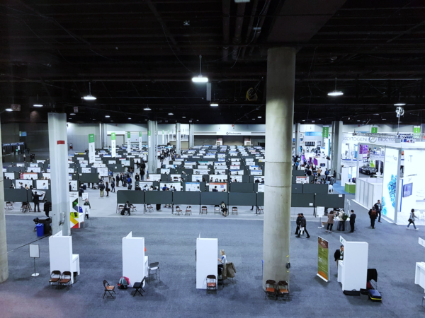
We are proud to be part of this new edition of AACR’23, which will take place in Orlando (Florida) from 14 to 19 April 2023!
The Oncodesign Services team will be present at the booth #1055 to exchange about our integrated Drug Discovery services in oncology.


Founded in 1907, the American Association for Cancer Research (AACR) is the first and largest research organization dedicated to accelerating the conquest of cancer.
Each year, the AACR Annual Meeting brings together scientists, clinicians, and other healthcare professionals to share the latest advances in cancer science and medicine.

Posters
Our scientists have prepared 5 posters to share our recent scientific developments at the meeting:
1
DRIVE-Biologics: All the steps from discovery to development of novel biological entities
- Session Date and Time: Monday Apr 17, 2023 9:00 AM – 12:30 PM
- Location: Poster Section 14
- Poster Board Number: 17
- Published Abstract Number: 1537
- Abstract:
Biologics first revolutionized cancer treatment in the late 1900s with the approval of rituximab and trastuzumab, two monoclonal antibodies targeting antigens expressed on tumor cells. Another milestone was achieved in the early 2010s with the approval of antibodies targeting immune checkpoints. Nowadays, the discovery and development of new biological entities and biological therapeutic products represent a rapidly growing market in various therapeutic areas, with about 10 to 15 biologics being approved each year. We have built a premium expert ecosystem services – DRIVE-Biologics – to support and accelerate biologics drug discovery and development in oncology, immuno-oncology, and inflammatory diseases. The DRIVE-Biologics consortium provides a unique integrated solution with specialist services from strategic partners, to access market analysis, establish the ability to design, optimize, and develop novel biological entities addressing the therapeutic target of interest. DRIVE-Biologics supplies the high-level, IND-focused discipline to rigorously manage the integrated programs integrating CMC, manufacturing, regulatory affairs and clinical trial. We will present the lead optimization and multiparameter preclinical evaluation process to select and assess biological candidates for downstream development and clinical studies:
– Custom cellular model development for discovery and potency analysis;
-In vitro screening, target engagement, and mechanism of action elucidation with cellular models ranging from tumor cell lines, immune cells or primary patient samples;
-In vivo efficacy and safety studies using refined and highly characterized syngeneic, xenogeneic, patient-derived xenograft or humanized mouse models up to non-human primates,
-DMPK capabilities to develop and validate bioanalytical methods (including GLP-compliancy) such as LBA and qPCR/RT-qPCR and also assess immunogenicity
-Biodistribution and tumor specificity analysis of bioconjugated and radiolabeled biologics. Herein, we provide DRIVE-BIO optimization and preclinical evaluation process to select promising biologics and list the key parameters to be checked. We will present our recent results which highlight the importance to optimize these parameters to improve the efficacy of biologics.
2
Simultaneous 18F-FDG PET / MR imaging for metastases identification in a disseminated human preclinical melanoma model
- Session Date and Time: Monday Apr 17, 2023 1:30 PM – 5:00 PM
- Location: Poster Section 17
- Poster Board Number: 21
- Published Abstract Number: 2767
- Abstract:
Melanoma is a skin cancer at high risk of metastatic progression, hence associated with poorer prognosis. Imaging represents an essential tool to assess tumor stage, but also to monitor disease progression and therapy response. However, a single imaging technique cannot efficiently detect and characterize all metastases. Therefore, we evaluated the potential of simultaneous 18F-FDG PET/MR imaging in preclinical models of disseminated melanoma at different stages.
Female Nude and SDRG rats were intravenously injected with CMEL 5 cells, originating from brain metastases of a human melanoma. On day (D) 60, 70, and 80 post-injection, Nude rats were imaged using brain T2-weighted (T2w) 7T MRI and whole-body simultaneous 18F-FDG (10-15 MBq) PET/MR. Furthermore, whole-body and brain T2w MR images were acquired using a 4.7T MRI on D20 and D34 post-injection in SDRG rats. Visual detection and quantitative analysis of lesions was performed on MRI or PET/MR images, and results were confronted with necropsy (melanin pigmented lesions) and gamma counting data.
From D60 post-CMEL-5 cell injection, all Nude animals exhibited metastases in one to four different locations. Moreover, most of metastases locations found in clinical setting were detected in our experimental model using simultaneous 18F-FDG PET/MR imaging, even allowing the observation of brain lesions on T2w MR images. MRI further allowed precise contouring of each organ of interest to effectively identify bone, lungs, adrenal and spleen metastases with enhanced 18F-FDG uptake. Additionally, PET image quantification in adrenals was consistent with gamma counting results. As anticipated, small lesions below PET spatial resolution limit (i.e. <1 mm) were not detected by imaging. SDRG rats displayed a faster and more homogenous tumor spread in brain, lungs and liver on MR images, while more metastases were seen during autopsy (additional spreading to pancreas, adrenals, reproductive system, bone, lymph nodes and skin).
The experimental model of disseminated CMEL-5 melanoma in Nude rats demonstrates its ability to mimic the human pathology. Hence, it appears to be a promising tool to monitor therapy response and potentially recognize progression from pseudo-progression. Simultaneous PET/MR imaging has shown to be more effective than stand-alone techniques in detecting melanoma dissemination, except for brain and small lesions for which the PET data are altered by partial volume effect. Thus, by complementing previously acquired MR images from the novel SDRG rat model with simultaneously acquired PET images, we could also provide insights on this promising metastatic model.
3
OncoTAM, a comprehensive preclinical platform to explore macrophages as key drivers of cancer progression and develop new therapies against tumor-associated-macrophages
- Session Date and Time: Tuesday Apr 18, 2023 9:00 AM – 12:30 PM
- Location: Poster Section 24
- Poster Board Number: 22
- Published Abstract Number: 4133
- Abstact :
Tumor-Associated Macrophages (TAMs) play an important role in the development of tumors, modulation of neoangiogenesis, immune suppression, and metastasis. A high infiltration of macrophages in the tumor is also correlated with a poor prognosis in several cancer types. Therefore, they became an attractive target for cancer immunotherapies. Several macrophage-targeting approaches in anticancer therapy are under development, including TAM depletion, inhibition of new TAM differentiation, or re-education of TAM activation for cancer cell phagocytosis. In this presentation, we will share different examples of in vitro assays and in vivo models we are implementing to support preclinical development of novel TAM-targeting strategies. Antibody-dependent cellular phagocytosis (ADCP) has been used to demonstrate one crucial mechanism of action of different antibody (Ab) therapies targeting macrophages. Some of these Ab (including anti-CSF1) have then been tested in syngeneic in vivo tumor models. To reach this goal, we demonstrated that the tumor implantation site in mice (subcutaneous vs orthotopic) could impact the polarization of macrophages (M1 vs M2). Differences in the ratio of M1 and M2 subtypes infiltrating the PAN-02 pancreatic murine tumors were observed, and anti-CSF1 antibodies increased the survival of mice bearing orthotopic Renca murine kidney tumor by eliminating TAMs. Additionally, in xenograft models of human breast tumors in NOD-SCID mice, the eradication of TAMs by anti-CSF1R clearly demonstrated the importance of macrophages in the tumor progression and in the anti-tumor efficacy of Abs mediated by macrophages. By using an orthotopic Hepa1-6 murine liver cancer model, we showed high antitumor efficacy of compounds targeting the STAT6 pathway by reprogramming immunosuppressive TAMs into an M1 phenotype that promotes the induction of a cytotoxic immune response. For compounds displaying no cross-reactivity with murine target, we developed models and characterized the TAMs in breast, colon, melanoma and head & neck PDX and CDX tumors in different huCD34-engrafted mouse models. Altogether, the panel of in vitro assays and in vivo tumor models in OncoTAM should be useful to provide insights on the mechanism of action and antitumor efficacy of novel immune-oncology strategies targeting macrophages.
4
Antitumor activity comparison of two somatostatin receptor ligands radiolabeled with Lutetium-177 (SSO110 and DOTA-TATE) alone or combined with chemotherapy in mice bearing AR42J SST2-positive tumors
- Session Date and Time: Tuesday Apr 18, 2023 1:30 PM – 5:00 PM
- Location: Poster Section 19
- Poster Board Number: 11
- Published Abstract Number: 5044
- Abstract :
Antitumor activity comparison of two somatostatin receptor ligands radiolabeled with Lutetium-177 (SSO110 and DOTA-TATE) alone or combined with chemotherapy in mice bearing AR42J SSTR2-Positive Tumors
Only a limited number of studies have compared the therapeutic effects of repetitive cycles of radiolabeled somatostatin (SST) analogues or assessed potential synergy of these analogues with chemotherapy agents.
Hence, a comparison was conducted in an in vivo AR42J tumor model with a SSTR2 antagonist, [177Lu]Lu-satoreotide tetraxetan ([177Lu]Lu‑SSO110), and a SSTR2 agonist, [177Lu]Lu-DOTA-TATE. The efficacy of treatment combining [177Lu]Lu-satoreotide tetraxetan with capecitabine plus temozolomide was also assessed.
Swiss Nude mice were xenografted with AR42J tumor cells. For the evaluation of the tumor response to radiation therapy, animals received one weekly dose of [177Lu]Lu‑SSO110 or [177Lu]Lu-DOTA-TATE for four consecutive weeks. For combined treatments, animals received one weekly dose of [177Lu]Lu‑SSO110 at 20 MBq for four consecutive weeks in association with daily doses of capecitabine and temozolomide (CAPTEM).
Weekly treatment for four consecutive weeks with [177Lu]Lu‑SSO110 at 15 MBq revealed a significantly reduced tumor growth, with 68 days to reach a tumor volume of 850 mm3, compared to [177Lu]Lu-DOTA-TATE at 15 MBq or 30 MBq, with, respectively, 43 days and 48 days. This was associated with a 3.5 fold higher tumor uptake of [177Lu]Lu‑SSO110 (15 MBq) compared to [177Lu]Lu-DOTA-TATE, with no or mild effects on body weight, hematological toxicity, or renal toxicity.
In AR42J tumor-bearing mice treated once a week for four consecutive weeks, [177Lu]Lu‑SSO110 (15 MBq) associated with CAPTEM regimen did not significantly increase the median time to reach a tumor volume of 1000 mm3 as compared to treatment with [177Lu]Lu-satoreotide alone: 50.7 vs 42.2 days, respectively. The association of the two treatments only led to mild weight losses.
Repeated administrations of [177Lu]Lu‑SSO110 were able to potentiate peptide receptor radionuclide therapy with a higher tumor uptake and longer median survival compared to [177Lu]Lu-DOTA-TATE. The The combination of [177Lu]Lu-satoreotide with capecitabine and temozolomide did not show any synergy, but no antagonism was observed either.
5
New oncology target identification and validation platform combining artificial intelligence and preclinical pharmacology
- Session Date and Time: Tuesday Apr 18, 2023 1:30 PM – 5:00 PM
- Location: Poster Section 31
- Poster Board Number: 19
- Published Abstract Number: 5370
- Abstract :
Despite major advances in cancer therapy in the last decades, treatment resistance can develop over time. Precision medicine allows for the successful implementation of targeted therapies and stratification of patients, but treatment resistance remains a major obstacle in patient management. The identification and validation of new targets associated with cancer resistance remains a major challenge. The great diversity of molecular mechanisms involved in treatment resistance phenomena, whether intrinsic (de novo or primary) or acquired (secondary), constitutes a real therapeutic challenge for patient care. A better understanding of resistance mechanisms would allow to explore new therapeutic strategies to circumvent these phenomena in different types of cancer. The OncoSNIPE® project was developed in this context as part of a multicenter and collaborative clinical study (NCT04548960) in more than 800 chemo-naive adult patients. The objective of this project was to identify early and/or late markers of treatment resistance in three different pathologies for which resistance problems are encountered: triple negative breast cancer (TNBC) or luminal B, locally advanced or metastatic non-small cell lung cancer (NSCLC) and pancreatic ductal adenocarcinoma (PDAC). The program included traditional clinical and whole exome sequencing (WES) monitoring of patient biopsies (Exom-seq and RNA-seq) at diagnosis and relapse, monitoring of blood markers (RNA-seq and Proteomics – Cytokine) at diagnosis, and the evaluation of best therapeutic responses and relapse. The program used bioinformatics, artificial intelligence, statistical learning and semantic enrichment approaches to discover the diversity of mechanisms involved in these resistances and to identify new therapeutic targets, through hetero-modal data including clinical, genomic, transcriptomic, immunological and radiomic dimensions. Subsequently, a specific flowchart for target validation was applied to the resulting list, considering the target’s developmental potential, its essentiality, prior knowledge (database mining) and home-made score of the link between the target and the disease. Finally, new targets were prioritized using weighting parameter and heuristic approximation based on the Crank algorithm. Experimental work on multiple targets began in the laboratory, initially in vitro, using 2D and 3D cell culture (including cells from patient-derived xenografts) and molecular interference. A wide variety of intrinsic or acquired molecular mechanisms involved in treatment resistance are being evaluated as candidates for diagnostic and therapeutic development.
- Session Date and Time: Monday Apr 17, 2023 9:00 AM – 12:30 PM
- Location: Poster Section 14
- Poster Board Number: 17
- Published Abstract Number: 1537
- Abstract:
Biologics first revolutionized cancer treatment in the late 1900s with the approval of rituximab and trastuzumab, two monoclonal antibodies targeting antigens expressed on tumor cells. Another milestone was achieved in the early 2010s with the approval of antibodies targeting immune checkpoints. Nowadays, the discovery and development of new biological entities and biological therapeutic products represent a rapidly growing market in various therapeutic areas, with about 10 to 15 biologics being approved each year. We have built a premium expert ecosystem services – DRIVE-Biologics – to support and accelerate biologics drug discovery and development in oncology, immuno-oncology, and inflammatory diseases. The DRIVE-Biologics consortium provides a unique integrated solution with specialist services from strategic partners, to access market analysis, establish the ability to design, optimize, and develop novel biological entities addressing the therapeutic target of interest. DRIVE-Biologics supplies the high-level, IND-focused discipline to rigorously manage the integrated programs integrating CMC, manufacturing, regulatory affairs and clinical trial. We will present the lead optimization and multiparameter preclinical evaluation process to select and assess biological candidates for downstream development and clinical studies:
– Custom cellular model development for discovery and potency analysis;
-In vitro screening, target engagement, and mechanism of action elucidation with cellular models ranging from tumor cell lines, immune cells or primary patient samples;
-In vivo efficacy and safety studies using refined and highly characterized syngeneic, xenogeneic, patient-derived xenograft or humanized mouse models up to non-human primates,
-DMPK capabilities to develop and validate bioanalytical methods (including GLP-compliancy) such as LBA and qPCR/RT-qPCR and also assess immunogenicity
-Biodistribution and tumor specificity analysis of bioconjugated and radiolabeled biologics. Herein, we provide DRIVE-BIO optimization and preclinical evaluation process to select promising biologics and list the key parameters to be checked. We will present our recent results which highlight the importance to optimize these parameters to improve the efficacy of biologics.
- Session Date and Time: Monday Apr 17, 2023 1:30 PM – 5:00 PM
- Location: Poster Section 17
- Poster Board Number: 21
- Published Abstract Number: 2767
- Abstract:
Melanoma is a skin cancer at high risk of metastatic progression, hence associated with poorer prognosis. Imaging represents an essential tool to assess tumor stage, but also to monitor disease progression and therapy response. However, a single imaging technique cannot efficiently detect and characterize all metastases. Therefore, we evaluated the potential of simultaneous 18F-FDG PET/MR imaging in preclinical models of disseminated melanoma at different stages.
Female Nude and SDRG rats were intravenously injected with CMEL 5 cells, originating from brain metastases of a human melanoma. On day (D) 60, 70, and 80 post-injection, Nude rats were imaged using brain T2-weighted (T2w) 7T MRI and whole-body simultaneous 18F-FDG (10-15 MBq) PET/MR. Furthermore, whole-body and brain T2w MR images were acquired using a 4.7T MRI on D20 and D34 post-injection in SDRG rats. Visual detection and quantitative analysis of lesions was performed on MRI or PET/MR images, and results were confronted with necropsy (melanin pigmented lesions) and gamma counting data.
From D60 post-CMEL-5 cell injection, all Nude animals exhibited metastases in one to four different locations. Moreover, most of metastases locations found in clinical setting were detected in our experimental model using simultaneous 18F-FDG PET/MR imaging, even allowing the observation of brain lesions on T2w MR images. MRI further allowed precise contouring of each organ of interest to effectively identify bone, lungs, adrenal and spleen metastases with enhanced 18F-FDG uptake. Additionally, PET image quantification in adrenals was consistent with gamma counting results. As anticipated, small lesions below PET spatial resolution limit (i.e. <1 mm) were not detected by imaging. SDRG rats displayed a faster and more homogenous tumor spread in brain, lungs and liver on MR images, while more metastases were seen during autopsy (additional spreading to pancreas, adrenals, reproductive system, bone, lymph nodes and skin).
The experimental model of disseminated CMEL-5 melanoma in Nude rats demonstrates its ability to mimic the human pathology. Hence, it appears to be a promising tool to monitor therapy response and potentially recognize progression from pseudo-progression. Simultaneous PET/MR imaging has shown to be more effective than stand-alone techniques in detecting melanoma dissemination, except for brain and small lesions for which the PET data are altered by partial volume effect. Thus, by complementing previously acquired MR images from the novel SDRG rat model with simultaneously acquired PET images, we could also provide insights on this promising metastatic model.
- Session Date and Time: Tuesday Apr 18, 2023 9:00 AM – 12:30 PM
- Location: Poster Section 24
- Poster Board Number: 22
- Published Abstract Number: 4133
- Abstact :
Tumor-Associated Macrophages (TAMs) play an important role in the development of tumors, modulation of neoangiogenesis, immune suppression, and metastasis. A high infiltration of macrophages in the tumor is also correlated with a poor prognosis in several cancer types. Therefore, they became an attractive target for cancer immunotherapies. Several macrophage-targeting approaches in anticancer therapy are under development, including TAM depletion, inhibition of new TAM differentiation, or re-education of TAM activation for cancer cell phagocytosis. In this presentation, we will share different examples of in vitro assays and in vivo models we are implementing to support preclinical development of novel TAM-targeting strategies. Antibody-dependent cellular phagocytosis (ADCP) has been used to demonstrate one crucial mechanism of action of different antibody (Ab) therapies targeting macrophages. Some of these Ab (including anti-CSF1) have then been tested in syngeneic in vivo tumor models. To reach this goal, we demonstrated that the tumor implantation site in mice (subcutaneous vs orthotopic) could impact the polarization of macrophages (M1 vs M2). Differences in the ratio of M1 and M2 subtypes infiltrating the PAN-02 pancreatic murine tumors were observed, and anti-CSF1 antibodies increased the survival of mice bearing orthotopic Renca murine kidney tumor by eliminating TAMs. Additionally, in xenograft models of human breast tumors in NOD-SCID mice, the eradication of TAMs by anti-CSF1R clearly demonstrated the importance of macrophages in the tumor progression and in the anti-tumor efficacy of Abs mediated by macrophages. By using an orthotopic Hepa1-6 murine liver cancer model, we showed high antitumor efficacy of compounds targeting the STAT6 pathway by reprogramming immunosuppressive TAMs into an M1 phenotype that promotes the induction of a cytotoxic immune response. For compounds displaying no cross-reactivity with murine target, we developed models and characterized the TAMs in breast, colon, melanoma and head & neck PDX and CDX tumors in different huCD34-engrafted mouse models. Altogether, the panel of in vitro assays and in vivo tumor models in OncoTAM should be useful to provide insights on the mechanism of action and antitumor efficacy of novel immune-oncology strategies targeting macrophages.
- Session Date and Time: Tuesday Apr 18, 2023 1:30 PM – 5:00 PM
- Location: Poster Section 19
- Poster Board Number: 11
- Published Abstract Number: 5044
- Abstract :
Antitumor activity comparison of two somatostatin receptor ligands radiolabeled with Lutetium-177 (SSO110 and DOTA-TATE) alone or combined with chemotherapy in mice bearing AR42J SSTR2-Positive Tumors
Only a limited number of studies have compared the therapeutic effects of repetitive cycles of radiolabeled somatostatin (SST) analogues or assessed potential synergy of these analogues with chemotherapy agents.
Hence, a comparison was conducted in an in vivo AR42J tumor model with a SSTR2 antagonist, [177Lu]Lu-satoreotide tetraxetan ([177Lu]Lu‑SSO110), and a SSTR2 agonist, [177Lu]Lu-DOTA-TATE. The efficacy of treatment combining [177Lu]Lu-satoreotide tetraxetan with capecitabine plus temozolomide was also assessed.
Swiss Nude mice were xenografted with AR42J tumor cells. For the evaluation of the tumor response to radiation therapy, animals received one weekly dose of [177Lu]Lu‑SSO110 or [177Lu]Lu-DOTA-TATE for four consecutive weeks. For combined treatments, animals received one weekly dose of [177Lu]Lu‑SSO110 at 20 MBq for four consecutive weeks in association with daily doses of capecitabine and temozolomide (CAPTEM).
Weekly treatment for four consecutive weeks with [177Lu]Lu‑SSO110 at 15 MBq revealed a significantly reduced tumor growth, with 68 days to reach a tumor volume of 850 mm3, compared to [177Lu]Lu-DOTA-TATE at 15 MBq or 30 MBq, with, respectively, 43 days and 48 days. This was associated with a 3.5 fold higher tumor uptake of [177Lu]Lu‑SSO110 (15 MBq) compared to [177Lu]Lu-DOTA-TATE, with no or mild effects on body weight, hematological toxicity, or renal toxicity.
In AR42J tumor-bearing mice treated once a week for four consecutive weeks, [177Lu]Lu‑SSO110 (15 MBq) associated with CAPTEM regimen did not significantly increase the median time to reach a tumor volume of 1000 mm3 as compared to treatment with [177Lu]Lu-satoreotide alone: 50.7 vs 42.2 days, respectively. The association of the two treatments only led to mild weight losses.
Repeated administrations of [177Lu]Lu‑SSO110 were able to potentiate peptide receptor radionuclide therapy with a higher tumor uptake and longer median survival compared to [177Lu]Lu-DOTA-TATE. The The combination of [177Lu]Lu-satoreotide with capecitabine and temozolomide did not show any synergy, but no antagonism was observed either.
- Session Date and Time: Tuesday Apr 18, 2023 1:30 PM – 5:00 PM
- Location: Poster Section 31
- Poster Board Number: 19
- Published Abstract Number: 5370
- Abstract :
Despite major advances in cancer therapy in the last decades, treatment resistance can develop over time. Precision medicine allows for the successful implementation of targeted therapies and stratification of patients, but treatment resistance remains a major obstacle in patient management. The identification and validation of new targets associated with cancer resistance remains a major challenge. The great diversity of molecular mechanisms involved in treatment resistance phenomena, whether intrinsic (de novo or primary) or acquired (secondary), constitutes a real therapeutic challenge for patient care. A better understanding of resistance mechanisms would allow to explore new therapeutic strategies to circumvent these phenomena in different types of cancer. The OncoSNIPE® project was developed in this context as part of a multicenter and collaborative clinical study (NCT04548960) in more than 800 chemo-naive adult patients. The objective of this project was to identify early and/or late markers of treatment resistance in three different pathologies for which resistance problems are encountered: triple negative breast cancer (TNBC) or luminal B, locally advanced or metastatic non-small cell lung cancer (NSCLC) and pancreatic ductal adenocarcinoma (PDAC). The program included traditional clinical and whole exome sequencing (WES) monitoring of patient biopsies (Exom-seq and RNA-seq) at diagnosis and relapse, monitoring of blood markers (RNA-seq and Proteomics – Cytokine) at diagnosis, and the evaluation of best therapeutic responses and relapse. The program used bioinformatics, artificial intelligence, statistical learning and semantic enrichment approaches to discover the diversity of mechanisms involved in these resistances and to identify new therapeutic targets, through hetero-modal data including clinical, genomic, transcriptomic, immunological and radiomic dimensions. Subsequently, a specific flowchart for target validation was applied to the resulting list, considering the target’s developmental potential, its essentiality, prior knowledge (database mining) and home-made score of the link between the target and the disease. Finally, new targets were prioritized using weighting parameter and heuristic approximation based on the Crank algorithm. Experimental work on multiple targets began in the laboratory, initially in vitro, using 2D and 3D cell culture (including cells from patient-derived xenografts) and molecular interference. A wide variety of intrinsic or acquired molecular mechanisms involved in treatment resistance are being evaluated as candidates for diagnostic and therapeutic development.