

Atopic dermatitis mouse models and pharmacology studies that support translational confidence.
Successful atopic dermatitis drug development requires more than demonstrating biological activity. Oncodesign Services has extensive experience designing and delivering preclinical pharmacology studies that combine clinically relevant models with comprehensive efficacy endpoints to provide a deeper understanding of patient-relevant therapeutic response.
Our translational approach helps generate robust preclinical evidence to better inform clinical decision-making, with bespoke model development available where standard models do not provide the best fit for your study.
Model design and selection
Atopic dermatitis (AD) is a chronic inflammatory skin disease characterized by recurrent eczematous lesions, intense pruritus, and dysregulation of immune and skin barrier functions. Robust preclinical models are essential for investigating disease mechanisms, evaluating therapeutic candidates, and identifying translational biomarkers.
Our dedicated auto-immunity and inflammation pharmacology team can help you select the best models to recapitulate the features of human atopic dermatitis you require, while keeping 3Rs best practice and animal welfare at the forefront of study design and implementation.
Our in vivo models bank for atopic dermatitis includes:
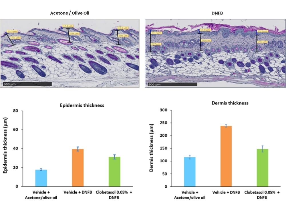
- DNFB-induced model in mice
- HDM-induced model in mice
- Calcipotriol/MC903-induced model in mice
- SpA-induced model (in development)
Combining different models and approaches enhances the translational relevance and robustness of preclinical research in atopic dermatitis drug development.
Confirm model suitability before study initiation: To support confident model selection, we also offer histology samples from established research models that can be accessed ahead of full study commitment to confirm feasibility. Immunohistochemistry (IHC) or FISH analyses may be performed to assess target expression and tissue localization, helping confirm biological relevance before program initiation. In atopic dermatitis, we offer samples from calcipotriol and DNFB models.
Our typical readouts for in vivo atopic dermatitis research:
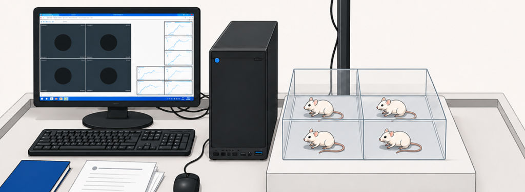
- Clinical scoring
- Real-time scratching
- Histopathology
- Biomarker / drug monitoring
- Genomics
Case studies and examples
-
House Dust Mite (HDM) Atopic Dermatitis Model
Topical application of HDM to BALB/c mice induces epidermis and dermis thickening, and recruitment of inflammatory cells (eosinophils, mast cells and CD4+ cells). HDM are applied directly on the skin and the area is bandaged. The bandage is regularly replaced over the course of several weeks.
This model is useful for testing the efficacy of compounds targeting general skin inflammation.
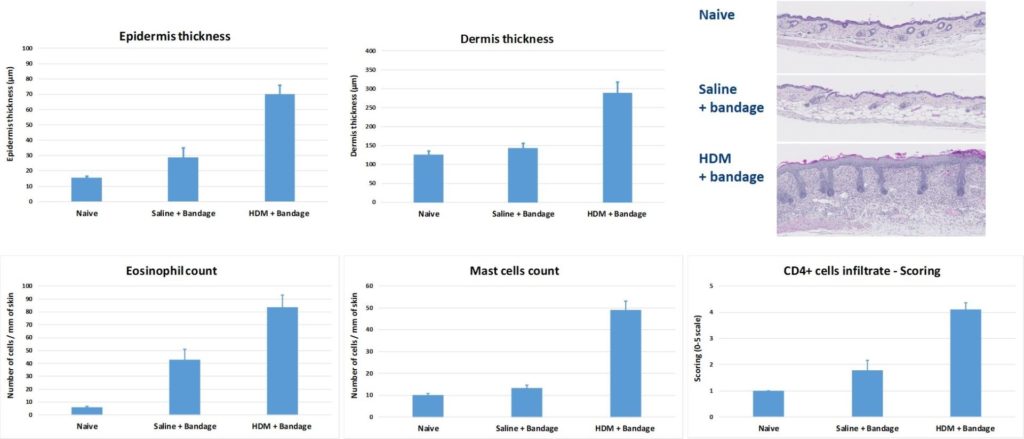
Induction of skin layer thickening, eosinophil, mast cell and CD4+ cell infiltration after HDM epicutaneous exposure.
-
Calcipotriol-induced Atopic Dermatitis in Mice
Topical application of Calcipotriol to BALB/c mice (BID from D0 to D9) induces changes in skin morphology and inflammation, resembling immune perturbations observed in acute lesions of atopic dermatitis in patients.
Topical drugs show protective effects on skin clinical score by reducing epidermis and dermis hyperplasia.
- Betamethasone (steroid anti-inflammatory drug)
- Ruxolinitib (JAK1/JAK2 inhibitor)
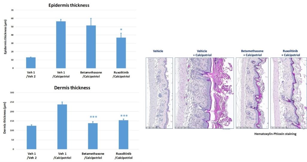
Left: Comparison of skin layer thickness with and without Calcipotriol application, and rescue by topical steroids or JAK2 inhibitor. Right: Representative histological images (H&P stain) obtained with and without calcipotriol application, and after rescue by topical steroids or JAK2 inhibitor.
Oncodesign Services (ODS) has been a trusted preclinical partner for several years. The team consistently demonstrates scientific excellence and reliability, tailoring preclinical protocols to our exact needs.
Their ability to rapidly action and deliver projects has made collaboration in oncology research efficient and straightforward. Clear, responsive communication is crucial to us, and the strong working relationship with our client manager makes coordination very easy.
We appreciate the professionalism, confidence, and flexibility ODS brings to every project.
Oncology Biopharma Organization (Europe)
Learn more about partnering with Oncodesign Services:
Oncodesign Services combines translational inflammation experience with a flexible approach to preclinical research, supporting both standard and customized study designs. Alongside an extensive portfolio of acute and chronic inflammatory disease models, we offer bespoke model development to address specific scientific questions and emerging therapeutic approaches.
Our scientific team are available to provide guidance from study planning through data interpretation, with comprehensive readouts including histology, clinical scoring, biomarkers, and functional endpoints. Histology samples are also available to support feasibility assessments and model selection before study initiation.
Contact our team to discuss your research objectives, explore the most appropriate models and endpoints, or request a quotation.

Frequently asked questions about atopic dermatitis models:
What biomarkers are typically evaluated in atopic dermatitis studies?
Frequently analyzed biomarkers include:
- IL-4, IL-13, IL-31, and TSLP
- Serum IgE
- Eosinophil infiltration
- Epidermal thickness
- Filaggrin expression
- TEWL (transepidermal water loss)
Biomarker selection is influenced by study objectives and model type. Speak with our inflammation team for more tailored information.
Can preclinical atopic dermatitis models be used for biologics and small molecules?
Yes, atopic dermatitis mouse models are widely used to evaluate:
- Monoclonal antibodies
- JAK inhibitors
- Small molecules
- Topical formulations
- RNA-based therapeutics
- Microbiome-targeted therapies
Study design can be adapted to the therapeutic modality.
How are itch or pruritus measured in research models?
Pruritus is commonly assessed by:
- Scratching frequency
- Scratching duration
- Neural and cytokine biomarker profiling
These endpoints help evaluate anti-pruritic therapies, as pruritus is a major hallmark of atopic dermatitis and among the most burdensome symptoms.
How do you select the right atopic dermatitis model for a study?
Model selection is influenced by factors such as:
- Therapeutic mechanism of action
- Desired endpoints
- Acute vs. chronic inflammation
- Barrier vs. immune focus
- Regulatory requirements
- Budget and timeline
A tailored strategy often provides the most informative data. Speak with our team to find out more.