Preclinical CRO Services for Asthma diseases
Developing treatments for asthma diseases involves the use of various models to study the underlying mechanisms and test potential therapeutic interventions.
Oncodesign Services offers access to several standard preclinical models addressing a variety of lung pathologies, including asthma, and provides CRO support for de novo development of new inflammation models recently described in literature.
Typical readouts for lung inflammation
Body weight
Lung weight
Clinical score
Edema
Antibody Production (specific IgG, IgE)
Mucus production
Lung histology
Gene expression
Ex vivo lung function, including compliance and elasticity (FlexiVent, SCIREQ)
Broncho-alveolar fluid (BALF) cell count and cytokine secretion
CT scan
Biomarker / drug monitoring
Oncodesign Services offers asthma models for your preclinical programs
Oncodesign Services, a CRO specialized in Inflammatory Diseases Research, helps you to choose the best models to recapitulate the features of human asthma disease and consider the ethical implications associated with animal research.
Our in vivo models bank includes:
- Allergic asthma model in mice, induced by House Dust Mice (HDM)
- Asthma exacerbation model in mice, induced by RSV + HDM combination
- (In development) Corticoid-resistant asthma in mice, induced by Ovalbumin (OVA) + Influenzae
Combining different models and approaches enhances the translational relevance and robustness of preclinical research in asthma drug development.
Case study : House dust-mite (HDM)-induced allergic asthma model
HDM is one of the most important sources of indoor allergens and a significant factor underlying allergic rhinitis and allergic asthma. HDM intranasal challenge can be used in BALB/c mice to reproduce many key features of clinical asthma:
- HDM challenge (IN) administered 5 days per week during 3 weeks
- Elevated levels of IgE
- Airway inflammation
- Goblet cell hyperplasia with mucus overproduction
- Release of inflammatory mediators and cytokines primarily associated with Th2-type inflammation
- Responsive to fluticasone (inhaled corticoid)
Results show allergic signature in bronchoalveolar lavage fluid (BALF) with eosinophilia in BALF & cytokine secretion, and in plasma with IgE levels.
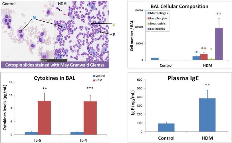
Allergic signature inbronchoalveolar lavage (BAL) and in plasma :
- BAL cell content & cytokine secretion
- Plasma IgE levels