

MRI, a powerful tool for preclinical oncology studies
MRI (Magnetic Resonance Imaging) is an imaging tool used in preclinical studies to optimize the homogeneity of randomization on tumor bearing mice and monitor the tumor volume during the course of a study. This allows you to add value to the preclinical data package while limiting the number of animals.
With 25 years of experience in Oncology, Oncodesign Services is the right partner for your preclinical programs supporting them by non-invasive imaging technologies such as MRI.
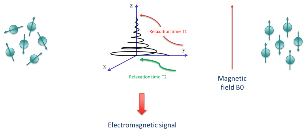
What are basic principles of MRI?
Magnetic Resonance Imaging (MRI) is a powerful non-invasive imaging technique that utilizes strong magnetic fields and radiofrequency pulses to generate detailed images of internal structures in living organisms.
In preclinical studies, MRI is commonly employed to investigate anatomy, physiology, and pathology in small animal models such as mice and rats. MRI can also support the evaluation of the mechanism of action of a drug (angiogenesis, metabolism…).
Preclinical MRI is a valuable tool in translational research, bridging the gap between basic research and clinical studies in humans. It allows scientists to non-invasively study a disease, evaluate treatment effects. Consequently, you gain a better understanding of biological processes in a controlled and ethical manner before moving to clinical trials.
-
Example of Morphological Magnetic Resonance Imaging
Morphological imaging applications:
- Anatomical imaging of soft tissues
- Non-invasives follow-up of tumor growth (figure1)
- Early detection of antitumor efficacy
- Use of contrast agents to enhance tissue signal
- Tumor localization and evaluation of disease progress (metastasis)
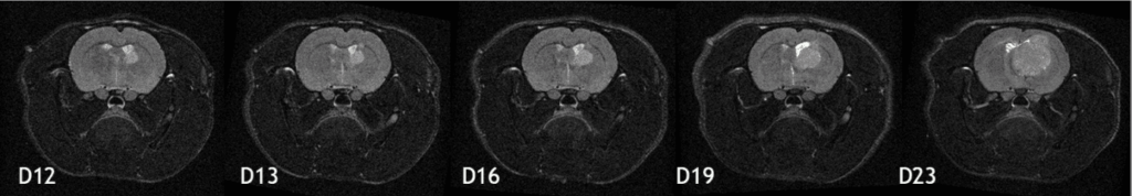
Figure 1: Serial monitoring of the growth of an orthotopically xenografted human glioblastoma (U-87 MG) bu T2-weighted MRI. Imaging timepoints in days post tumor implantation
MRI capabilities:
- Baseline image acquisition using also contract agent (e.g Gadolinium))
- Dynamic follow-up of uptake in the tumor
- Quantitative and standardized image analysis using in-house developed software
- Analyzed parameters:
- Initial Area Under the Curve (iAUC)
- Ktrans tumor mapping
- Control / Validation with gold standard immune-histochemistry assays :
- Semi-quantitative analysis to evaluate neo-angionesis and vessel maturity (CD31/SMA co-staining)
- Functional assay to measure the number and size of vessels and to characterize vascular permeability (Hoechst/Dextran FITC)
-
Example of Dynamic Contrast-Enhanced Magnetic Resonance Imaging (DCE-MRI)
Applications :
- Characterize tumor perfusion and microvascular permeability
- Support proof of concept / mechanism of action
- Optimize biological active dose, scheduling in combination therapies
- Identify biomarkers of efficacy for anti-angiogenic or anti-vascular drugs (Figure 2)
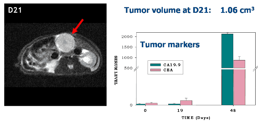
Figure 2: Earlier evaluation of tumor growth by MRI in combination to blood biomarkers in Nude rats bearing orthotopic BxPC-3 human pancreatic tumors.
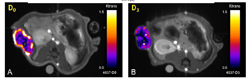
 (C)
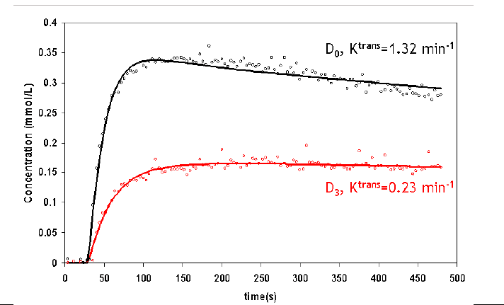
(C)Figure 3: Results from DCE-MRI experiment performed on Nude Rats bearing MDA-231 human breast tumor xenografts and treated from D0 with Sorafenib. Krans parameter maps superimposed on morphological images before (A) and after (B) treatment with corresponding uptake curves (C), showing the effect of Sorafenib on vascular status.
With an optimized workflow, morphological MRI is cost efficient, more ethical and adds value to your efficacy studies in preclinical models
Using common models (including MBT-2 bladder, U87 glioma, BT474 breast metastases, liver, prostate and others), Oncodesign Services has a deep experience in these advance translational models.