


Understand where your candidate distributes and how its distribution changes over time.
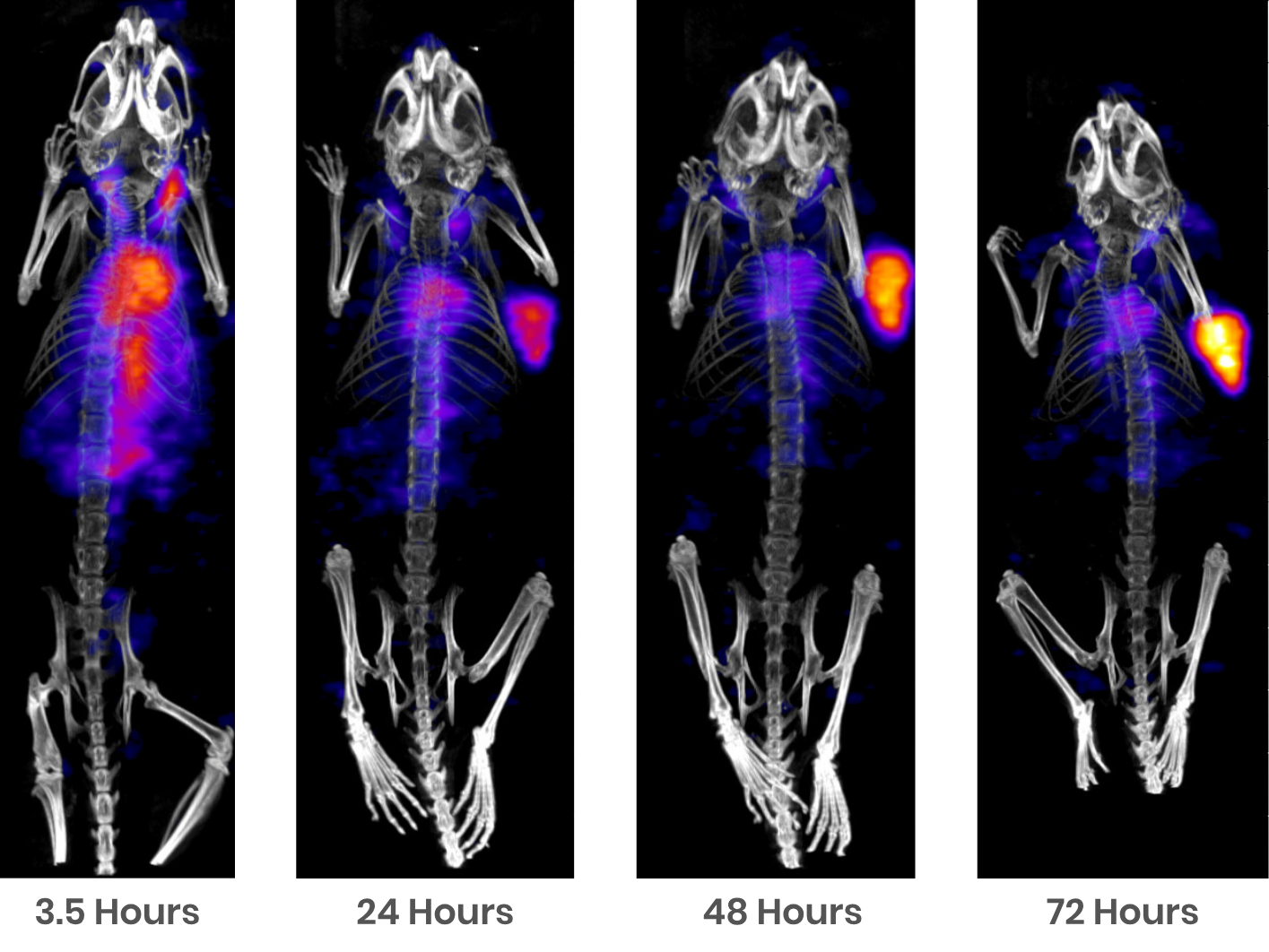
Biodistribution studies assess whether a candidate reaches its target, how long it persists and where off-target exposure may raise safety concerns. Blood measurements and endpoint tissue analyses provide important exposure data; however, longitudinal in vivo imaging provides whole-body data that evolves over time within the same animals, helping fully characterize tissue-associated signal while supporting reduction and refinement.
We provide comparative screening and detailed characterization biodistribution studies using a range of imaging methodologies including fluorescence, bioluminescence, PET, SPECT, CT and MRI.
Why Use Imaging for Biodistribution?
Imaging adds valuable information on where signals occur in the body and how they evolve between tissue collection time points, enabling researchers to:
- Increase statistical power of study data and reduce experimental variability: By monitoring the same models over time instead of relying on periodic endpoint collection of tissues from separate cohorts, the influence of between-animal variability on experimental data is drastically reduced, in addition to a significant reduction in the number of models required for a study.
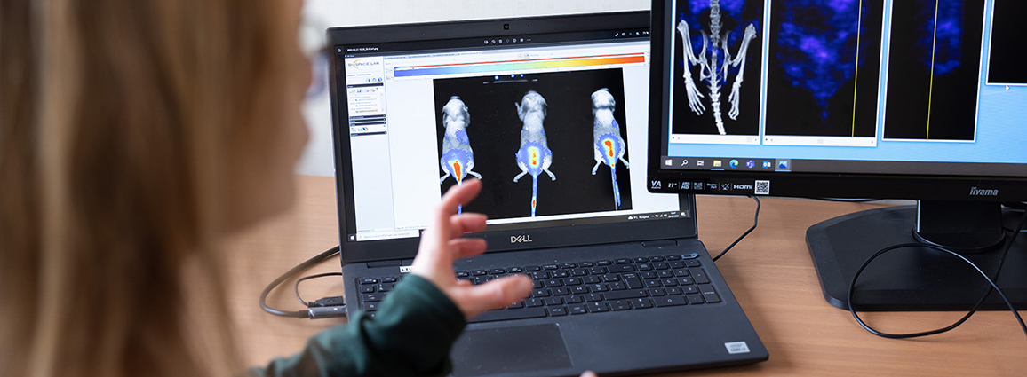
- Follow distribution over time: Assess uptake, retention and clearance longitudinally through repeated measurements in the same models. For luciferase-expressing cells or biological systems, monitor bioluminescent signals over time to assess their location and persistence, accounting for the reporter’s requirements and the biological conditions that influence light production.
- Support reduction and refinement: Repeated non-invasive imaging significantly reduces reliance on separate terminal cohorts.
- Visualize whole-body distribution: Suitable imaging approaches allow signals in multiple anatomical regions to be assessed within the same non-terminal examination. PET or SPECT combined with anatomical imaging helps localize uptake within organs and tissues.
- Compare candidates and doses: Examine distribution differences between compounds, biological constructs and doses. Comparative screening can help prioritize candidates for more extensive characterization.
- Connect imaging with tissue-level evidence: Combine whole-body observations with ex vivo imaging of collected tissues, gamma counting or other relevant assays. These complementary measurements help investigate signals that are difficult to distinguish in vivo.
Imaging Methods for Biodistribution
The appropriate imaging method depends on the material being studied, the tissue of interest, expected kinetics and the required level of quantification. Our imaging team assesses labeling or reporter compatibility before validating the acquisition and analysis strategy.
-
Optical Imaging
Fluorescence imaging
Compare fluorescently labeled compounds or doses and follow their distribution at selected time points. Imaging of organs can help distinguish tissue signals that overlap or are attenuated in whole-body scans.
Our fluorescence biodistribution studies typically include:
- Fluorophore selection and conjugation strategy.
- Assessment of biological properties after labeling.
- Detection feasibility and imaging setup.
- Longitudinal in vivo fluorescence imaging.
- Ex vivo organ imaging and more detailed tissue localization.
Fluorescence measurements are generally semi-quantitative in this setting. Tissue depth, autofluorescence and fluorophore properties influence the observed signal.
Bioluminescence imaging
Follow luciferase-expressing cells or biological systems through repeated in vivo imaging. Applications at Oncodesign Services include cell tracking and longitudinal assessment of viral-vector-mediated reporter expression.
Study preparation establishes suitable conditions for detecting and comparing signals. This typically includes:
- Evaluation of the reporter and substrate combination.
- Substrate dose and signal-kinetics assessment.
- Selection of acquisition timing.
- Longitudinal whole-body or region-focused imaging.
- Complementary imaging of collected tissues.
Bioluminescence reflects reporter activity. Interpretation must account for enzymatic reaction, substrate availability, tissue attenuation and acquisition conditions. Residual signal in case of repeated imaging and detector saturation also require consideration when comparing measurements.
-
Nuclear Imaging
PET and SPECT imaging
PET and SPECT imaging can be used to measure radiotracer uptake and distribution over time, with CT or MRI providing anatomical context. Study formats range from focused candidate comparisons to detailed biodistribution characterization.
Our nuclear imaging studies typically include:
- Isotope, chelator and linker selection.
- Bioconjugation and radiolabeling procedure development.
- Relevant in vitro assays and label-stability assessment.
- Longitudinal PET or SPECT imaging.
- Endpoint tissue radioactivity measurements via gamma counting.
Isotope half-life, biological kinetics, acquisition requirements and radionuclide supply guide study planning. Biodistribution data can also support radiopharmaceutical dosimetry studies.
Comparative Screening or Full Characterization?
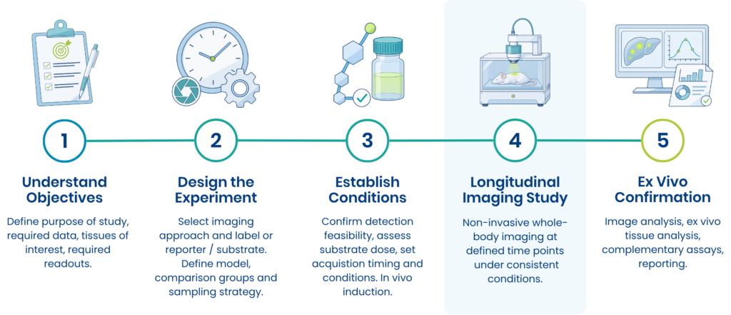
Imaging-based biodistribution studies can fulfill different purposes, depending on where you are in your drug discovery program. The number of candidates, imaging time points and endpoint measurements required for a biodistribution study are all typically influenced by the data required to support your next steps.
| Study Consideration | Comparative Screening | Full Biodistribution Characterization |
| Typical research phase | Hit-to-lead. | Lead optimization. |
| Primary objective | Identify distribution differences and prioritize candidates for further evaluation. | Characterize uptake, retention and clearance of selected candidates. |
| Imaging schedule | A focused set of time points selected to reveal relevant differences. | Multiple time points aligned with expected biological kinetics. |
| Candidate comparison | Compare compounds, variants or doses under a defined protocol. | Examine fewer candidates in greater depth. |
| Tissue confirmation | Selected tissues relevant to the screening objective. | A tissue panel selected to address the characterization objectives. |
| Example readouts | Relative distribution, tissue-associated signal and relevant signal ratios. Confirmation via ex vivo tissue analysis. | Longitudinal imaging measurements with complementary ex vivo tissue analysis. |
Biodistribution study structure
Discuss Your Biodistribution Requirements
Whether you need to compare candidates or investigate distribution in greater detail, our imaging and pharmacology scientists can help you review options and understand how imaging data can strengthen your preclinical data.

Frequently asked questions about imaging-based biodistribution studies:
How do I choose the right imaging method?
Selection depends on the candidate or biological system, whether labeling or reporter expression is feasible, the tissues of interest and the required readout. Fluorescence can support comparative assessment of labeled compounds. Bioluminescence supports studies of reporter-expressing biological systems. PET and SPECT provide quantitative radiotracer measurements.
When should biodistribution be investigated?
An early study can identify distribution constraints before extensive candidate optimization. The appropriate timing depends on the program, available material and whether sufficient biological evidence is available to justify in vivo evaluation.
Can you work with an already labeled compound or reporter-expressing system?
Existing conjugates and reporter-expressing systems can be assessed for compatibility and detection feasibility. When discussing this with our team, useful information includes label or reporter identity, biological activity, expected signal levels and the tissue of interest.
Can imaging replace ex vivo endpoint tissue analysis?
While non-invasive longitudinal imaging studies can drastically reduce the number of mice required for an in vivo biodistribution study, imaging and ex vivo tissue measurements provide complementary information. Longitudinal imaging follows signals within the same models, while endpoint measurements can confirm tissue distribution or investigate the biological material associated with the signal. The need for endpoint analysis depends on the study objective and detection strategy.
Does a low bioluminescence signal mean the biological system is absent?
A low or undetectable signal may reflect limited reporter expression, substrate availability, tissue attenuation or the detection limit. Acquisition optimization and complementary measurements help distinguish these possibilities.