



Lung inflammation models for respiratory disease and fibrosis research.
Oncodesign Services provides established respiratory inflammation models for asthma, asthma exacerbation, RSV-induced bronchiolitis, and pulmonary fibrosis. Studies can integrate inflammatory, histological, biomarker and functional endpoints, including BAL analysis, lung imaging and FlexiVent assessment of lung functionality.
Our scientists help you align the most appropriate model with induction protocol, treatment schedule and endpoints to meet your study objectives, with bespoke model development available where standardized models are not the best fit.
Typical readouts for respiratory disease models:
- Body weight
- Lung weight
- Lung weight-to-body weight ratio
- Clinical scoring
- Histology:
- Edema
- Fibrosis score
- Mucus production
- Leukocyte infiltration
- Viral load (for RSV)
- Lung function, including lung capacity, hyperesthesis, compliance and elasticity (FlexiVent, SCIREQ)
- Broncho-alveolar fluid (BALF) cell count and cytokine concentration
- CT scan
- Gene expression in lungs, by RT-qPCR / dPCR
- Biomarker / drug monitoring
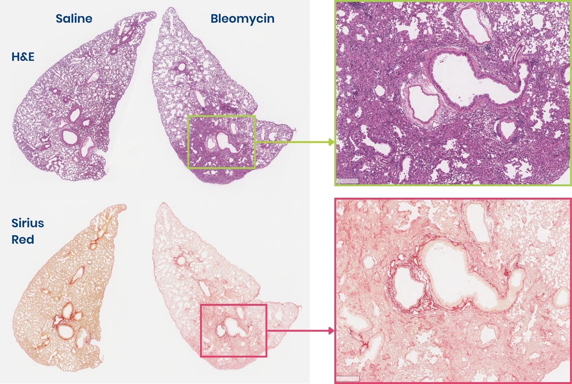
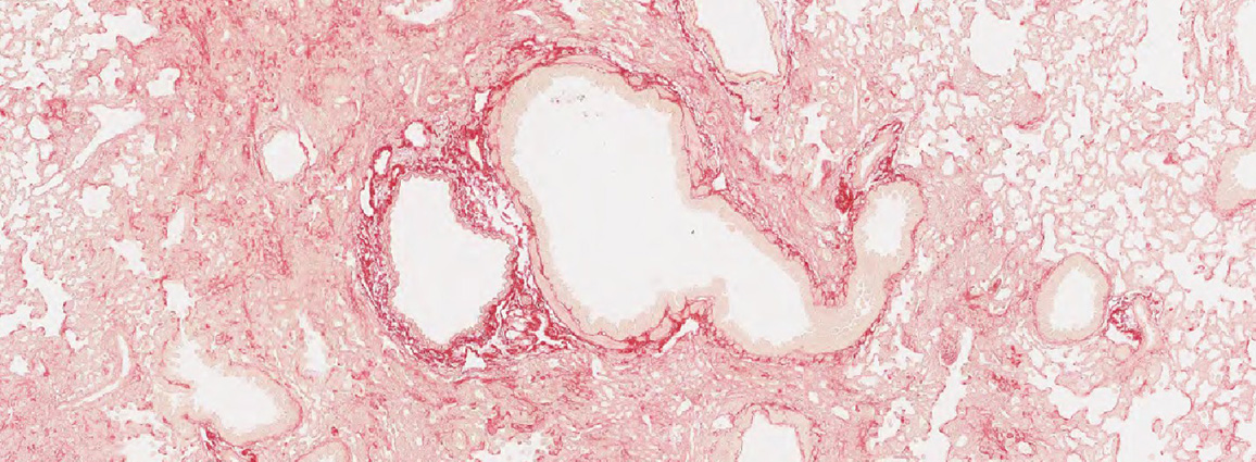
Histological assessment of lung fibrosis. H&E staining highlights inflammation and morphology (top). Sirus red staining highlights collagen deposition (bottom). Control on the left vs bleomycin-induced fibrosis on the right.
Common drug administration routes (PO, IV, IP, SC) and non-typical drug administration routes used for respiratory disease therapies:
- Intra-tracheal
- Intra-nasal
- Osmotic mini-pumps
Confirm feasibility before study initiation
To support confident model selection, histology samples from established respiratory disease models can be selectively accessed ahead of full study commitment. Immunohistochemistry (IHC) or FISH analyses may be performed to assess target expression and tissue localization, helping confirm biological relevance before program initiation.
Currently available lung inflammation samples include:
- HDM asthma
- Intratracheal bleomycin
Established lung inflammation models available for preclinical studies:
-
Pulmonary Fibrosis (IPF)
We offer bleomycin-induced pulmonary fibrosis models using intratracheal administration or delivery by subcutaneous osmotic minipumps. These approaches produce different lesion patterns and disease kinetics, supporting model selection according to the research question, with readouts including CT imaging, histology, gene expression and lung function.
- Lung fibrosis model in mice, induced by intratracheal administration of bleomycin
- Lung fibrosis model in mice, induced by bleomycin delivered through osmotic minipumps
-
Asthma
We offer mouse models of allergic asthma induced by house dust mite or ovalbumin, alongside an HDM–RSV exacerbation model. These models support efficacy assessment across allergic airway inflammation and infection-driven exacerbation, with readouts including BALF analysis, IgE, histology, gene expression and lung function.
- Allergic asthma model (2) in mice, induced by House Dust Mites (HDM)
- Allergic asthma model in mice, induced by Ovalbumin (OVA) sensitization
- Asthma exacerbation model (4) in mice, induced by RSV + HDM combination
-
Bronchiolitis
We offer a bronchiolotis model using a high titer of the human virus RSV, responsible for bronchiolitis, to infect mice. This model can generate viral replication in the lungs of mice, and supports the evaluation of antiviral and anti inflammatory therapies, with readouts including BALF analysis, viral titers, histology, and cytokine quantification.
- Bronchiolitis model (1) in mice, induced by Human Respiratory Syncytial Virus (RSV)
Experimental examples for respiratory disease studies
-
Asthma induction breakdown for the HDM allergy model
House dust mites (HDM) are one of the most common sources of indoor allergens and a significant factor underlying allergic rhinitis and allergic asthma. HDM intranasal challenge can be used in BALB/c mice to reproduce many key features of clinical asthma, including:
- Elevated levels of IgE
- Airway inflammation
- Goblet cell hyperplasia with mucus overproduction
- Release of inflammatory mediators and cytokines primarily associated with Th2-type inflammation
- Responsive to fluticasone (inhaled corticoid)
(HDM challenge (IN) administered 5 days per week over 3 weeks.)
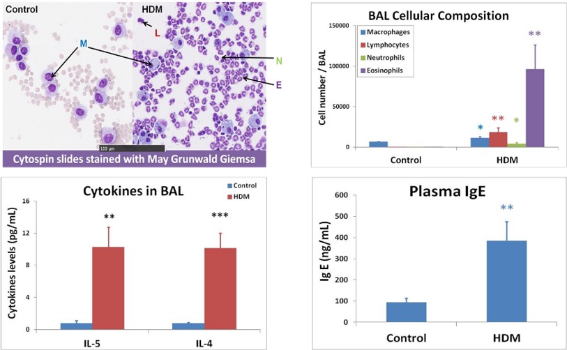
Results show allergic signature in bronchoalveolar lavage fluid (BALF) and in plasma with elevated eosinophil contents as well as TH2 cytokine concentrations in the BALF, and elevated IgE levels in the plasma.
-
Bronchiolitis induction via respiratory syncytial virus (RSV) in BALB/c mice
Respiratory syncytial virus (RSV) is a virus responsible for severe respiratory symptoms including rhinitis, bronchiolitis and pneumonia in young children. About 65% of children are infected with RSV within the first year of life.
Bronchiolitis is modeled by delivering a high titer of RSV by intranasal instillation to BALB/c mice, which allows viral infection and bronchiolitis symptoms. Excessive production of airway mucus is a feature of bronchiolitis and is a key histology readout. Ribavirin is typically used as a reference compound.
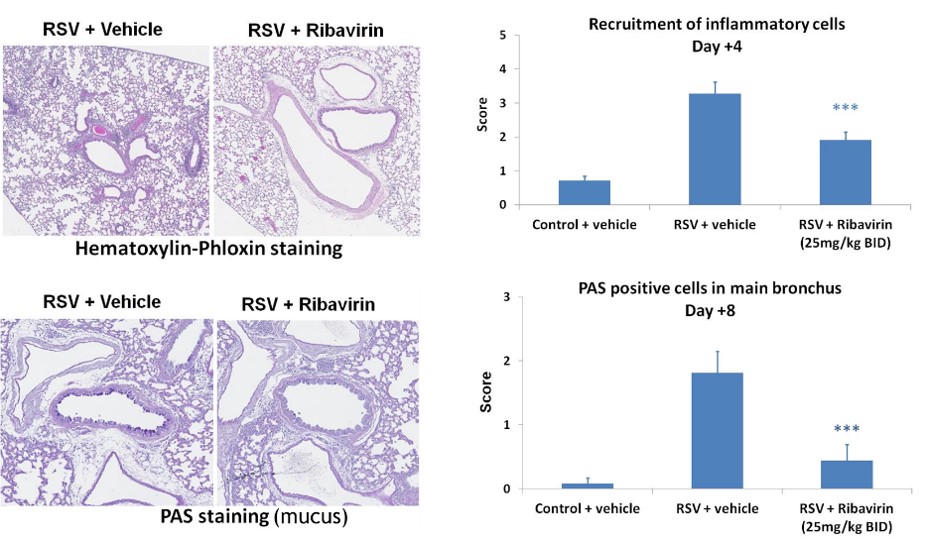
Histological modifications following RSV infection with and without Ribavirin treatment.
-
Induction of lung fibrosis via bleomycin using intra-tracheal instillation
The bleomycin-induced lung fibrosis mouse model is one of the most frequently used animal models for the investigation of pulmonary fibrosis resulting from inflammation.
Like the human disease, bleomycin exposure in mice is associated with epithelial damage, inflammatory cell infiltration, and proliferation of fibroblasts and myofibroblasts, as well as extra-cellular matrix excessive deposition.
Protocol: intra-tracheal (IT) administration of bleomycin on day 0 in C57BL/6 male mice (n=8 animals/group as a minimum after randomization).
- D0: Single IT administration of bleomycin or saline under ketamine/xylazine anaesthesia
- D7: Selection of good responders to bleomycin based on % body weight loss (selection rate of 65%)
- D7 to D21: Treatment
- Around D21: Endpoint studies
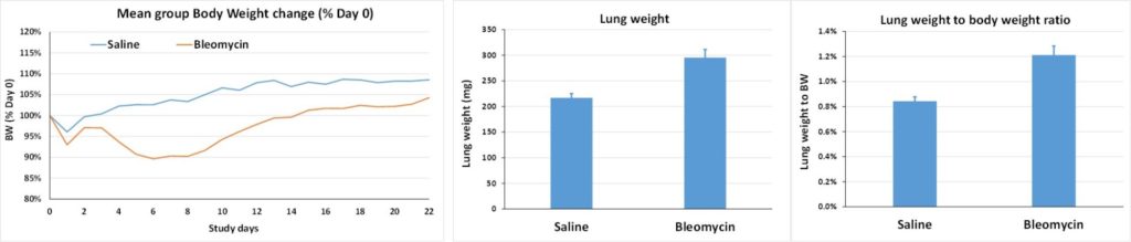
Effect of bleomycin induction compared to sham challenge with saline on body weight (Bleomycin group in orange, saline in blue, % of weight compared to D0, left), lung weight on D21 (middle) and lung weight on body weight ratio on D21 (right).
Lung histology (evaluation of lung total collagen content) on D21 for anti-fibrotic drug evaluation:
- ALK5 (TGFb receptor I) inhibitor [SB-525334]: effective at 30 or 60mg/kg/day (with Cmax-driven dose-effect)
- Nintedanib (Tyr kinase inhibitor): no effect at 60 or 100mg/kg/day
- Pirfenidone (molecular target unknown): no effect at 300 or 500mg/kg/day
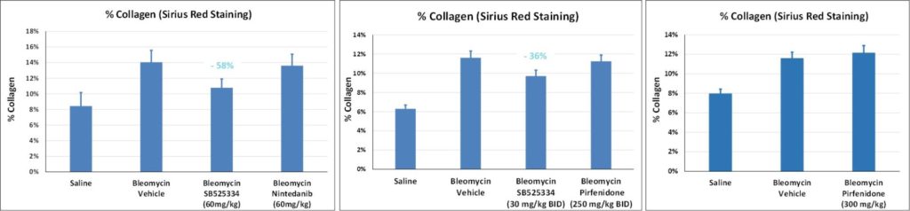
Comparative effect of SB525334 (ALK5 inhibitor), Pirfenidone and Nintedanib on collagen deposition in lungs (histological assessment, Sirius red) in the Bleomycin-induced lung fibrosis model (data from 3 separate studies).
Learn more about partnering with Oncodesign Services:
Oncodesign Services combines translational inflammation experience with a flexible approach to preclinical research, supporting both standard and customized study designs. Alongside an extensive portfolio of acute and chronic inflammatory disease models, we offer bespoke model development to address specific scientific questions and emerging therapeutic approaches.
Our scientific team is available to provide guidance from study planning through data interpretation, with comprehensive readouts including histology, clinical scoring, biomarkers, and functional endpoints. Histology samples are also available to support feasibility assessments and model selection before study initiation.
Contact our team to discuss your research objectives, explore the most appropriate models and endpoints, or request a quotation.
Oncodesign Services (ODS) has been a trusted preclinical partner for several years. The team consistently demonstrates scientific excellence and reliability, tailoring preclinical protocols to our exact needs.
Their ability to rapidly action and deliver projects has made collaboration in oncology research efficient and straightforward. Clear, responsive communication is crucial to us, and the strong working relationship with our client manager makes coordination very easy.
We appreciate the professionalism, confidence, and flexibility ODS brings to every project.
Oncology Biopharma Organization (Europe)



Frequently asked questions about our respiratory disease models:
Can Oncodesign Services customize respiratory disease studies?
Yes. Study designs can typically be customized to align with therapeutic targets, dosing strategies, biomarkers, endpoints, and timelines. Our teams may tailor challenge agents, readouts, imaging methods, and sample collection protocols to fit sponsor requirements.
Do you support biomarker and translational analyses?
Yes. Many preclinical lung studies incorporate translational biomarkers to help bridge preclinical and clinical findings. These may include cytokine panels, transcriptomics, flow cytometry, multiplex assays, and pharmacodynamic biomarkers.
Can you evaluate biologics and advanced therapeutics?
Yes. Lung inflammation models are frequently used to assess:
- Monoclonal antibodies
- Cell and gene therapies
- RNA-based therapeutics
- Small molecules
- Inhaled therapeutics
- Immunomodulators
-
References
(1) Bronchiolitis model in mice, induced by Human Respiratory Syncytial Virus (RSV)
Borchers AT, Chang C, Gershwin ME, Gershwin LJ. Respiratory syncytial virus–a comprehensive review. Clin Rev Allergy Immunol. 2013 Dec;45(3):331-79. doi: 10.1007/s12016-013-8368-9. PMID: 23575961; PMCID: PMC7090643.
https://pubmed.ncbi.nlm.nih.gov/23575961/
https://link.springer.com/article/10.1007/s12016-013-8368-9
Taylor G. Animal models of respiratory syncytial virus infection. Vaccine. 2017 Jan 11;35(3):469-480. doi: 10.1016/j.vaccine.2016.11.054. Epub 2016 Nov 29. PMID: 27908639; PMCID: PMC5244256.
https://www.ncbi.nlm.nih.gov/pmc/articles/PMC5244256/
Ceneviva ZJ, Norlander AE, Stokes Peebles R Jr. Mouse Models of Respiratory Syncytial Virus Infection. Methods Mol Biol. 2022;2506:19-41. doi: 10.1007/978-1-0716-2364-0_2. PMID: 35771461.
https://pubmed.ncbi.nlm.nih.gov/35771461/
Tregoning JS, Yamaguchi Y, Harker J, Wang B, Openshaw PJ. The role of T cells in the enhancement of respiratory syncytial virus infection severity during adult reinfection of neonatally sensitized mice. J Virol. 2008 Apr;82(8):4115-24. doi: 10.1128/JVI.02313-07. Epub 2008 Feb 13. PMID: 18272579; PMCID: PMC2293007.
https://pubmed.ncbi.nlm.nih.gov/18272579/
Sebina I, Phipps S. The Contribution of Neutrophils to the Pathogenesis of RSV Bronchiolitis. Viruses. 2020 Jul 27;12(8):808. doi: 10.3390/v12080808. PMID: 32726921; PMCID: PMC7472258.
https://pubmed.ncbi.nlm.nih.gov/32726921/
(2) Allergic asthma model in mice, induced by House Dust Mice (HDM)
Gao X, Leung TF, Wong GW, Ko WH, Cai M, He EJ, Chu IM, Tsang MS, Chan BC, Ling J, Fan X, Lu L, Lam CW, Wong CK. Meteorin-β/Meteorin like/IL-41 attenuates airway inflammation in house dust mite-induced allergic asthma. Cell Mol Immunol. 2022 Feb;19(2):245-259. doi: 10.1038/s41423-021-00803-8. Epub 2021 Nov 30. PMID: 34848868; PMCID: PMC8803866.
https://pubmed.ncbi.nlm.nih.gov/34848868/
Debeuf N, Haspeslagh E, van Helden M, Hammad H, Lambrecht BN. Mouse Models of Asthma. Curr Protoc Mouse Biol. 2016 Jun 1;6(2):169-184. doi: 10.1002/cpmo.4. PMID: 27248433.
https://pubmed.ncbi.nlm.nih.gov/27248433/
Oikonomou N, Schuijs MJ, Chatzigiagkos A, Androulidaki A, Aidinis V, Hammad H, Lambrecht BN, Pasparakis M. Airway epithelial cell necroptosis contributes to asthma exacerbation in a mouse model of house dust mite-induced allergic inflammation. Mucosal Immunol. 2021 Sep;14(5):1160-1171. doi: 10.1038/s41385-021-00415-5. Epub 2021 May 27. PMID: 34045680; PMCID: PMC8379077.
https://pubmed.ncbi.nlm.nih.gov/34045680/
Ma M, Li G, Qi M, Jiang W, Zhou R. Inhibition of the Inflammasome Activity of NLRP3 Attenuates HDM-Induced Allergic Asthma. Front Immunol. 2021 Aug 3;12:718779. doi: 10.3389/fimmu.2021.718779. PMID: 34413860; PMCID: PMC8369415.
https://pubmed.ncbi.nlm.nih.gov/34413860/
Maes B, Smole U, Vanderkerken M, Deswarte K, Van Moorleghem J, Vergote K, Vanheerswynghels M, De Wolf C, De Prijck S, Debeuf N, Pavie B, Toussaint W, Janssens S, Savvides S, Lambrecht BN, Hammad H. The STE20 kinase TAOK3 controls the development of house dust mite-induced asthma in mice. J Allergy Clin Immunol. 2022 Apr;149(4):1413-1427.e2. doi: 10.1016/j.jaci.2021.08.020. Epub 2021 Sep 8. PMID: 34506849.
https://pubmed.ncbi.nlm.nih.gov/34506849/
(3) Corticoid-resistant asthma in mice, induced by Ovalbumin (OVA) + Influenzae
Essilfie AT, Simpson JL, Dunkley ML, Morgan LC, Oliver BG, Gibson PG, Foster PS, Hansbro PM. Combined Haemophilus influenzae respiratory infection and allergic airways disease drives chronic infection and features of neutrophilic asthma. Thorax. 2012 Jul;67(7):588-99. doi: 10.1136/thoraxjnl-2011-200160. Epub 2012 Mar 3. PMID: 22387445.
https://pubmed.ncbi.nlm.nih.gov/22387445/
Kim RY, Pinkerton JW, Essilfie AT, Robertson AAB, Baines KJ, Brown AC, Mayall JR, Ali MK, Starkey MR, Hansbro NG, Hirota JA, Wood LG, Simpson JL, Knight DA, Wark PA, Gibson PG, O’Neill LAJ, Cooper MA, Horvat JC, Hansbro PM. Role for NLRP3 Inflammasome-mediated, IL-1β-Dependent Responses in Severe, Steroid-Resistant Asthma. Am J Respir Crit Care Med. 2017 Aug 1;196(3):283-297. doi: 10.1164/rccm.201609-1830OC. PMID: 28252317.
https://pubmed.ncbi.nlm.nih.gov/28252317/
(4) Asthma exacerbation model in mice, induced by RSV + HDM combination
Zhang D, Yang J, Zhao Y, Shan J, Wang L, Yang G, He S, Li E. RSV Infection in Neonatal Mice Induces Pulmonary Eosinophilia Responsible for Asthmatic Reaction. Front Immunol. 2022 Feb 2;13:817113. doi: 10.3389/fimmu.2022.817113. PMID: 35185908; PMCID: PMC8847141.
https://pubmed.ncbi.nlm.nih.gov/35185908/
https://www.frontiersin.org/articles/10.3389/fimmu.2022.817113/full
Makino A, Shibata T, Nagayasu M, Hosoya I, Nishimura T, Nakano C, Nagata K, Ito T, Takahashi Y, Nakamura S. RSV infection-elicited high MMP-12-producing macrophages exacerbate allergic airway inflammation with neutrophil infiltration. iScience. 2021 Oct 2;24(10):103201. doi: 10.1016/j.isci.2021.103201. PMID: 34703996; PMCID: PMC8524145.
https://pubmed.ncbi.nlm.nih.gov/34703996/
Matsuse H, Hirose H, Tsuchida T, Fukahori S, Fukushima C, Mizuta Y, Kohno S. Effects of respiratory syncytial virus infection on dendritic cells and cysteinyl leukotrienes in lung tissues of a murine model of asthma. Allergol Int. 2007 Jun;56(2):165-9. doi: 10.2332/allergolint.O-06-476. Epub 2007 May 1. PMID: 17460444.
https://pubmed.ncbi.nlm.nih.gov/17460444/
(5) Lung fibrosis model in mice, induced by Bleomycin delivered IT or by osmotic pumps
Herrmann FE, Hesslinger C, Wollin L, Nickolaus P. BI 1015550 is a PDE4B Inhibitor and a Clinical Drug Candidate for the Oral Treatment of Idiopathic Pulmonary Fibrosis. Front Pharmacol. 2022 Apr 20;13:838449. doi: 10.3389/fphar.2022.838449. PMID: 35517783; PMCID: PMC9065678.
https://www.frontiersin.org/articles/10.3389/fphar.2022.838449/full
Lee R, Reese C, Bonner M, Tourkina E, Hajdu Z, Riemer EC, Silver RM, Visconti RP, Hoffman S. Bleomycin delivery by osmotic minipump: similarity to human scleroderma interstitial lung disease. Am J Physiol Lung Cell Mol Physiol. 2014 Apr 15;306(8):L736-48. doi: 10.1152/ajplung.00210.2013. Epub 2014 Feb 28. PMID: 24583879; PMCID: PMC3989726.
https://pubmed.ncbi.nlm.nih.gov/24583879/
Liang M, Lv J, Zou L, Yang W, Xiong Y, Chen X, Guan M, He R, Zou H. A modified murine model of systemic sclerosis: bleomycin given by pump infusion induced skin and pulmonary inflammation and fibrosis. Lab Invest. 2015 Mar;95(3):342-50. doi: 10.1038/labinvest.2014.145. Epub 2014 Dec 15. PMID: 25502178.
https://www.nature.com/articles/labinvest2014145
Hübner RH, Gitter W, El Mokhtari NE, Mathiak M, Both M, Bolte H, Freitag-Wolf S, Bewig B. Standardized quantification of pulmonary fibrosis in histological samples. Biotechniques. 2008 Apr;44(4):507-11, 514-7. doi: 10.2144/000112729. PMID: 18476815.
https://pubmed.ncbi.nlm.nih.gov/18476815/
Egger C, Gérard C, Vidotto N, Accart N, Cannet C, Dunbar A, Tigani B, Piaia A, Jarai G, Jarman E, Schmid HA, Beckmann N. Lung volume quantified by MRI reflects extracellular-matrix deposition and altered pulmonary function in bleomycin models of fibrosis: effects of SOM230. Am J Physiol Lung Cell Mol Physiol. 2014 Jun 15;306(12):L1064-77. doi: 10.1152/ajplung.00027.2014. Epub 2014 Apr 11. PMID: 24727584.
https://pubmed.ncbi.nlm.nih.gov/24727584/
De Langhe E, Cailotto F, De Vooght V, Aznar-Lopez C, Vanoirbeek JA, Luyten FP, Lories RJ. Enhanced endogenous bone morphogenetic protein signaling protects against bleomycin induced pulmonary fibrosis. Respir Res. 2015 Mar 15;16(1):38. doi: 10.1186/s12931-015-0202-x. PMID: 25849157; PMCID: PMC4364322.
https://pubmed.ncbi.nlm.nih.gov/25849157/
Dackor RT, Cheng J, Voltz JW, Card JW, Ferguson CD, Garrett RC, Bradbury JA, DeGraff LM, Lih FB, Tomer KB, Flake GP, Travlos GS, Ramsey RW Jr, Edin ML, Morgan DL, Zeldin DC. Prostaglandin E₂ protects murine lungs from bleomycin-induced pulmonary fibrosis and lung dysfunction. Am J Physiol Lung Cell Mol Physiol. 2011 Nov;301(5):L645-55. doi: 10.1152/ajplung.00176.2011. Epub 2011 Aug 19. PMID: 21856819; PMCID: PMC3213994.
https://pubmed.ncbi.nlm.nih.gov/21856819/
Phillips JE, Peng R, Burns L, Harris P, Garrido R, Tyagi G, Fine JS, Stevenson CS. Bleomycin induced lung fibrosis increases work of breathing in the mouse. Pulm Pharmacol Ther. 2012 Aug;25(4):281-5. doi: 10.1016/j.pupt.2011.10.001. Epub 2011 Oct 20. Erratum in: Pulm Pharmacol Ther. 2022 Jun;73-74:102122. PMID: 22024054.
https://pubmed.ncbi.nlm.nih.gov/22024054/