


Metastasis models supported by pharmacology and preclinical imaging for the investigation of new metastatic cancer therapies.
For most cancer patients, a primary, solid tumor can be treated with surgery. However, refractory reoccurrence and metastasis remain common among cancer patients, with a significant rate of mortality. For this reason, metastasis models are an essential part of the preclinical oncology toolkit.
Oncodesign Services offers a range of metastasis models that vary by injection site and address the complex metastatic cascade, from the generation of circulating tumor cells (CTCs) to tumor growth at a distal site.
Tumor burden and dissemination of metastatic tumors can be monitored via longitudinal in vivo imaging including MRI, PET/MRI, PET, SPECT and optical imaging, and via endpoint ex vivo examination.
Metastasis models:
Metastasis models with intact immune system (Syngeneic)
| Primary Pathology | Cell Lines | Model System | Inoculation Route | Injection Site | Metastatic Site | Incidence Rate | Standard of Care |
| Breast | 4T1 | Balb/c | OT | MFP | Lung | 100% | Doxorubicin, Indomethacin |
| Colon | CT-26 | Balb/c | IV | N/A | Lung | 100% | N/A |
| Kidney | RENCA | Balb/c | OT | Kidney | Lung | 100% | Sunitinib, Sorafenib |
| Melanoma | B16-F10 | C57BL/6 | Intracranial IV | N/A | Brain | 100% | Temozolomide |
| Lung | 100% | Cyclophosphamide | |||||
| Sarcoma | NCTC 2472 | C3H/HeJ | Intraosseous | Intra-tibial | Bone | 100% | CDDP |
Metastasis models in immune-compromised mice
Here is the complete second table with the same full-width layout, padding and alternating shading. I’ve preserved its data and rowspan structure.
| Primary Pathology | Cell Lines | Model System | Inoculation Route | Injection Site | Metastatic Site | Incidence Rate | Standard of Care |
| Breast | BT-474 | Balb/c Nude | Intracranial | N/A | Brain | 100% | Paclitaxel CDDP Temozolomide Gemcitabine |
| MDA-MB-231 | Nude | IV | N/A | Lung, Lymph nodes | 66% | Paclitaxel | |
| NSG | OT | MFP | Lung | 50-75% | N/A | ||
| 4T1* | NSG | OT | MFP | Lung | 100% | Indomethacin | |
| Colon | LS 174T | Swiss Nude | Intrasplenic | N/A | Liver | 60-80% | CPT-11 |
| HCT-116 | CB17 SCID | OT | Caecum | Liver | 90% | CPT-11 | |
| Lung | A-549 | Nude Rat | Intracranial | N/A | Brain | 100% | N/A |
| NCI-H460 | Swiss Nude | Intracranial | N/A | Brain | 100% | Paclitaxel | |
| Melanoma | CMEL-5 | Swiss Nude | IV | N/A | Lymph node, Bone | 100% | Temozolomide |
| B16-F10* | Swiss Nude | OT | Skin | Lymph node | 100% | N/A | |
| Nude Rat | IV | N/A | Lymph node, Bone, Brain | 100% | N/A | ||
| Swiss Nude | Intradermal | Ear | Lung, Lymph node | 80-90% | N/A | ||
| Prostate | PC-3 | Nude Rat | Intracardiac | N/A | Bone, Lymph nodes, Adrenal glands | 69% | Paclitaxel |
| 22RV1 | NSG Mice | Intra-arterial | N/A | Bone, Liver | 90-100% | N/A | |
| Pancreas | AsPC-1 | BRGSF | Intrasplenic | Pancreas | Liver, Lungs | 100% | NA |
*Murine cell line engrafted in an immunocompromised strain.
Metastasis models in super-immune-compromised mice (PDX)
| Primary Pathology | Model | Model System | Inoculation Route | Primary Tumor Site | Metastatic Site | Incidence Rate | Standard of Care |
| Breast | OD-BRE-0192 | NSG | OT | MFP | Lung | 30% | N/A |
| Colon | CR-LRB-018P | SCID | OT | Caecum | Liver | 40% | CPT-11 |
| NOG | OT | Caecum | Liver | 100% | Sorafenib | ||
| Uvea melanoma | MP55 | TK-NOG Liver Humanized | OT | Choroid | Liver | 33% | N/A |
| Pancreas | IM-PAN-015 | NSG | SC | N/A | Lung | 40–60% | N/A |
| IM-PAN-010 | NSG | SC | Liver | 40% | |||
| OT | Liver | 40% |
Case studies and examples:
-
Testing Cyclophosphamide as a metastatic inhibitor in a B16-F10 melanoma model
The B16-F10 syngeneic melanoma model can be adapted to investigate primary tumor growth, therapeutic response and metastatic disease in immunocompetent C57BL/6 mice. Studies at Oncodesign Services have evaluated how implantation site, treatment timing and route of inoculation can be selected according to the experimental objective.
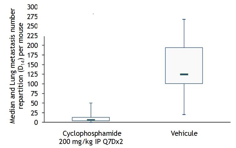
Demonstrated here, the B16-F10 melanoma model was implanted intravenously with lung tumor colonies measured at endpoint. Cyclophosphamide, a chemotherapy, successfully inhibited significant lung metastasis, when compared with the control:
For primary tumor studies, comparison of subcutaneous (SC) and mammary fat pad (MFP) implantation showed progressive tumor growth with both approaches, while MFP implantation significantly reduced tumor necrosis compared with the SC model.
The SC model has been characterized across multiple therapeutic modalities, including immune checkpoint inhibitors, STING agonists, chemotherapy and fractionated radiotherapy. Importantly, studies with anti-PD-1 and anti-CTLA-4 showed different responses depending on whether treatment began before or after an established tumor microenvironment was present, demonstrating how treatment timing can influence observed efficacy. Radiotherapy studies also demonstrated a dose-dependent response, while chemotherapy studies captured outcomes including tumor growth inhibition, relapse following treatment cessation and treatment-associated changes in body weight.
For metastatic research, intravenous inoculation can be used to establish lung metastases, with metastatic burden assessed through macroscopic analysis and qPCR. This configuration has been used both to investigate immune mechanisms involved in metastatic control and to evaluate therapeutic response. NK-cell depletion increased lung metastases, while Cyclophosphamide reduced metastatic burden compared with control.
Together, these data demonstrate the flexibility of the B16-F10 model and the importance of aligning implantation route, treatment timing and endpoints with the specific biological and therapeutic question being investigated.
-
Comparing intra-tibial and intra-arterial metastasis models for prostate cancer
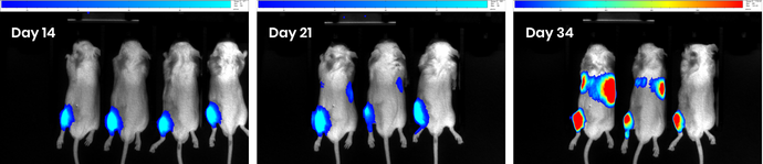
Researchers at Oncodesign Services compared two approaches for modelling metastatic prostate cancer in immunodeficient male mice. 22Rv1-Luc-mCherry human prostate cancer cells were administered by intra-tibial or intra-arterial injection, with longitudinal bioluminescence imaging used to characterize the timing and anatomical distribution of disease progression.
- Intra-tibial injection: Bioluminescence signals remained localized to the tibia for the first 21 days. From day 28, distant signals became detectable, with ex vivo imaging confirming dissemination to the liver and lungs.
- Intra-arterial injection: Early signals were observed in the bones, followed by increasingly prominent liver involvement. Endpoint imaging also identified signals in the hind-limb bone, spinal column and seminal vesicle.
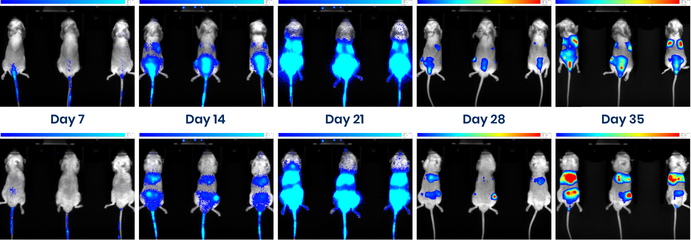
The two injection routes produced distinct patterns and kinetics of metastatic dissemination. This demonstrates that administration route influences the disease trajectory reproduced by a metastasis model.
In this study, longitudinal imaging provided information that endpoint analysis alone could not capture, including when dissemination became detectable and how metastatic burden developed over time. These data can help inform treatment initiation, cohort allocation and response assessment.
Learn more about partnering with Oncodesign Services:
Oncodesign Services brings over 30 years of oncology experience to preclinical studies. We provide both standard and customized research models, flexible pharmacology studies, and longitudinal preclinical imaging. We are fully AAALAC accredited.
Our scientific team is available to provide guidance from study planning through data interpretation, with comprehensive readouts including tumor burden (e.g. histology, imaging etc), clinical scoring, and biomarkers.
Contact our team to discuss your research objectives, explore the most appropriate models and endpoints, and request a quotation.
What do our clients say about us?
Working with Oncodesign Services has been a true drug development partnership – delivering great results over a period of years.
As an early-stage company, we needed a partner both we and our investors could trust to deliver on time and on budget as part of our regulatory submissions. Oncodesign’s oncology expertise and ability to bring together in vitro and in vivo work under one roof gave us that confidence from the outset. They also played an important role in coordinating assay method development and supporting external specialist laboratories working on radio-labelled materials and drug stability, performance studies, helping to connect biological findings with chemical and physical data.
Beyond the science, they have been an exceptional and reliable partner. From supporting our Brain Tumour Charity grant submission to co-presenting our work on MTL-004 at the AACR Annual Meeting in both 2024 and 2025, their contribution has always been consistent and meaningful.
ODS does exactly what it says it will do, on time and on budget, which is critical for a small company like ours.
Gordian Pharma Ltd



Frequently asked questions about our cancer metastasis models:
When should a metastasis model be included in a preclinical oncology program?
A metastasis model may be valuable when a therapeutic candidate targets a pathway involved in metastatic progression, or when evidence of activity beyond the primary tumor could help differentiate the candidate. Although metastatic studies are not required for every oncology program, they can provide additional evidence in disease settings where metastatic progression is clinically important.
How do I choose the most appropriate metastasis model for my candidate?
Model selection should reflect the candidate’s mechanism of action, the stage of the metastatic process being investigated, the expected site of dissemination and the intended study endpoints. The tumor model, implantation or injection route, host background and monitoring strategy should therefore be considered together.
Our oncology specialists are experienced in evaluating research briefs and designing studies with the most appropriate protocols to align with your objectives.
What is the difference between spontaneous and experimental metastasis models?
Spontaneous metastasis models begin with a primary tumor and allow tumor cells to progress through multiple stages of the metastatic cascade. Experimental metastasis models introduce tumor cells directly into the circulation or a secondary site, enabling more focused and reproducible assessment of later stages such as organ colonization and metastatic growth.
How does the injection route affect a metastasis model?
The injection route influences where tumor cells travel, which stages of metastasis are represented and where secondary tumors develop. Intravenous, intracardiac, intra-arterial, orthotopic and direct intra-organ approaches can therefore produce different disease patterns and answer different experimental questions.
How can metastatic tumor development be monitored?
Metastatic tumor burden and dissemination can be assessed using longitudinal in vivo imaging and endpoint analyses. Depending on the model and study objectives, monitoring may include bioluminescence imaging, MRI, PET, PET/MRI, SPECT or optical imaging, supported by ex vivo examination of relevant organs.