

Comparing histological outcomes with inspiratory capacity, static compliance and work of breathing.
Preclinical efficacy in pulmonary fibrosis is often demonstrated through changes in lung histology. Reduced collagen deposition, improvements in tissue architecture, and lower fibrosis scores can provide important evidence that a therapeutic intervention is having an effect. However, the patient experience is focused around lung functionality.
Idiopathic pulmonary fibrosis (IPF) is a chronic, progressive disease characterized by lung scarring, increasing tissue stiffness and impaired oxygen transfer. As the disease progresses, patients experience shortness of breath, persistent cough and fatigue. The act of breathing typically becomes increasingly difficult.
This creates an important translational question for preclinical researchers: If a treatment improves the histological signs of fibrosis, does lung function systematically improve with it? In other words, can preclinical projects be satisfied with improved histology, or should lung function and mechanics assessments be introduced to de-risk progression to clinical trials?
Data from a bleomycin-induced model of pulmonary fibrosis show why assessing different dimensions of therapeutic efficacy can provide a more complete picture of treatment response. They also highlight an important limitation of relying on histology alone to predict functional improvement.
In this article:
1. The difference between preclinical endpoints and clinical outcomes
2. Histological assessment of an ALK5 inhibitor with strong biological activity
3. Functional assessment, comparisons and correlations
4. Selecting endpoints for preclinical pulmonary fibrosis studies
5. Key takeaways
The difference between preclinical endpoints and clinical outcomes.
In clinical research for idiopathic pulmonary fibrosis, lung function is a major consideration. Forced vital capacity is widely used to assess disease progression and therapeutic response, alongside outcomes including hospitalization and mortality. Exercise tolerance, dyspnea and quality of life can provide further information about how disease affects patients.
Preclinical pulmonary fibrosis research tends to focus on histological endpoints, with good reason. Histology can provide valuable information about disease severity, tissue morphology and the extent of collagen deposition. Established scoring systems, including the modified Ashcroft score, allow the severity of pulmonary fibrosis to be evaluated and compared across experimental groups.
However, histology and lung function do not measure the same impact. Histology assesses changes in tissue structure, and lung function assesses what those changes mean for the respiratory function of the lungs. Understanding the relationship between the two can therefore be important when evaluating whether a therapeutic effect is likely to extend beyond changes in disease pathology into symptomatic benefit for a patient.
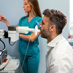
In the clinic, lung function in pulmonary fibrosis is a primary readout and commonly assessed using forced vital capacity (FVC), which measures the maximum volume of air a patient can forcibly exhale after a forced inspiration. In preclinical studies, the analogous measure is inspiratory capacity (IC), which quantifies the maximum volume of air the lungs can inhale after a normal exhalation. Although the techniques differ, both readouts assess changes in lung capacity associated with collagen deposition and therapeutic response.
In preclinical studies, inspiratory capacity is measured using specialized pulmonary function equipment that precisely controls and records the movement of air into the lungs. This allows researchers to quantify changes in lung capacity associated with fibrosis and determine whether a treatment preserves or restores respiratory function beyond what can be inferred from histology alone.
Assessment of an ALK5 inhibitor with strong biological activity.
Histological assessment:
In this study, a bleomycin-induced model of pulmonary fibrosis was used to assess SB-525334, a selective inhibitor of ALK5. ALK5 is the TGF-β type I receptor that plays a key role in the development of fibrosis. Following intratracheal administration of bleomycin, the models developed established pulmonary fibrosis characterized by inflammation, collagen deposition and changes in lung morphology.
Bleomycin substantially increased collagen deposition and modified Ashcroft scores:
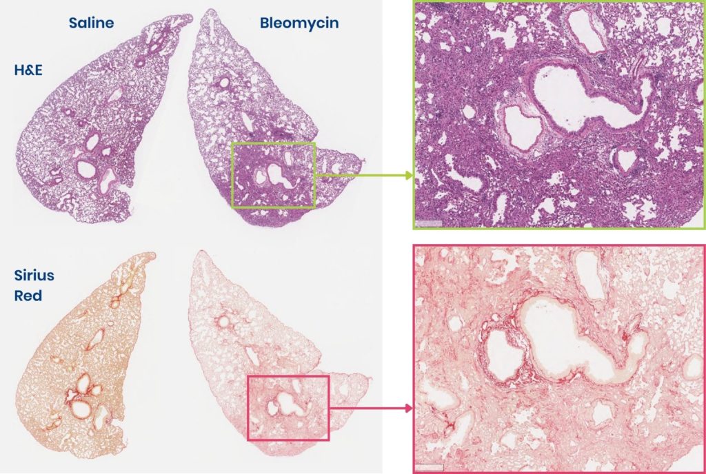
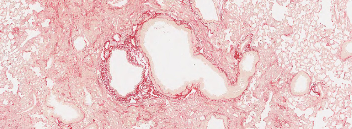
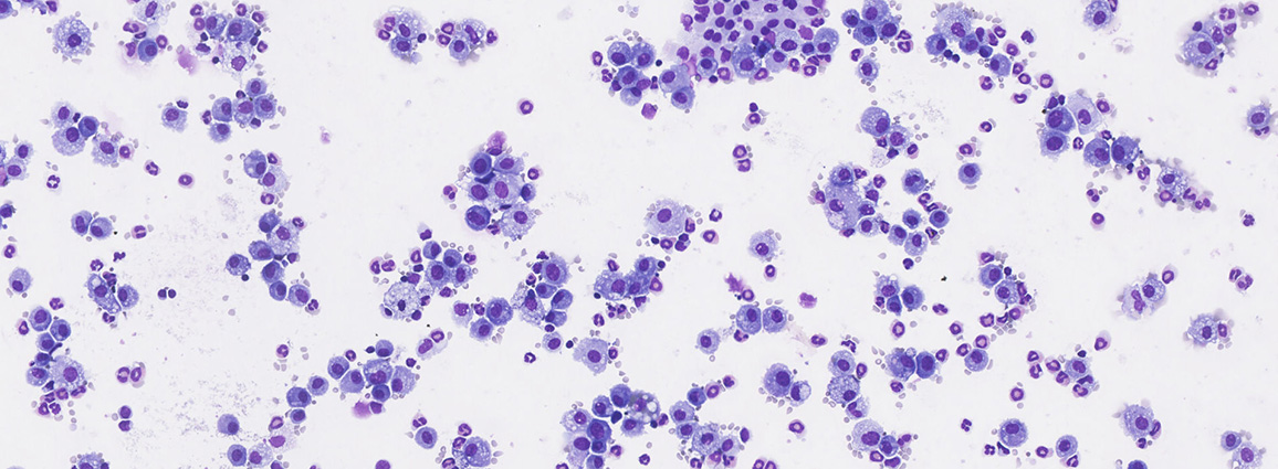
Caption: D21 histological assessment of lungs. H&E staining highlights inflammation and morphology (top). Sirus red staining highlights collagen deposition (bottom). Control (saline) on the left. Following single intratracheal administration of bleomycin on the right. Following bleomycin administration, we can see peribronchial inflammation and fibrosis in the lung tissue.
The mice were then treated with SB-525334 between day 7 and day 20-21. Histological assessment showed a clear treatment effect, with reduced collagen deposition and Ashcroft scores in the mice receiving the inhibitors compared to bleomycin alone.
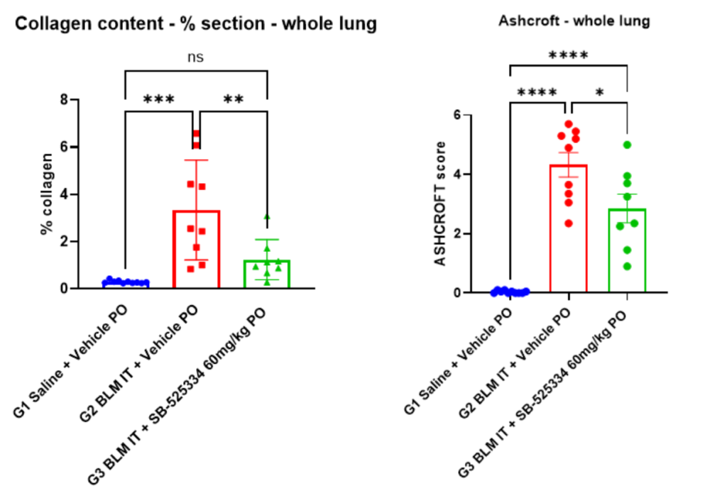
Caption: As we can see here, treatment with SB-525334 showed a significant reduction in collagen content (left) on D21 after D7-21 treatment, and a reduction in Ashcroft scoring (right).
If efficacy had been assessed through histology alone, the results would present convincing evidence of anti-fibrotic activity. The compound clearly demonstrates an impact on lung fibrosis; however, this is not a holistic picture of the extent to which impaired lung function has been recovered. When functional data was added, it was discovered that the magnitude of functional recovery was generally more moderate than the histological response.
Functional assessment, comparisons and correlations:
Lung function is not a single measurement. Different parameters capture different aspects of respiratory physiology and can therefore provide different information about disease progression and therapeutic response.
When performing preclinical lung function assessments, three parameters are informative: inspiratory capacity, static compliance, and normalized work of breathing. Here we compare the parameter with the functional and histological results from the SB-525334 study:
| Readout | Findings for SB-525334 | Evidence |
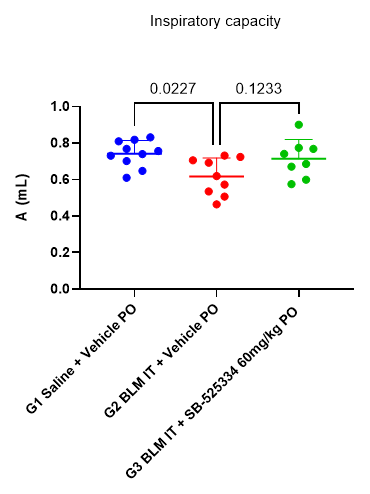
| Inspiratory capacity Inspiratory capacity describes the volume of air that can be inhaled following a normal expiration. |
In mice receiving bleomycin, inspiratory capacity decreased (red), compared with the control group (blue), reflecting the reduced ability of fibrotic lungs to accommodate air. Treatment with the ALK5 inhibitor produced a trend towards improvement (green), with the functional recovery being relatively modest. Histological assessment of the proportion of air space within the lung tissue showed a similar pattern. However, the relationship between histological air space and directly measured inspiratory capacity was not strong enough to suggest that one readout could reliably substitute for the other. |
 |
| Static compliance Static compliance measures the ability of lung tissue to expand. Healthy lungs are relatively compliant. As fibrosis develops and the tissue becomes stiffer, greater force is required to inflate the lungs and compliance decreases.Static compliance is typically the parameter that shows the highest and most sensitive response to fibrotic intervention. |
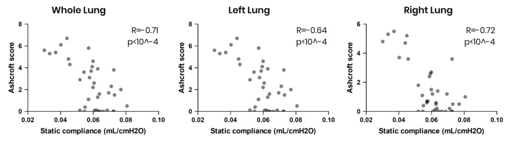
In the bleomycin model, static compliance was substantially reduced (red). Treatment with the ALK5 inhibitor produced a trend towards functional improvement (green). Among the functional parameters assessed, static compliance showed a particularly clear response to the induction of fibrosis. This is consistent with its frequent use as a functional endpoint in experimental pulmonary fibrosis research. Again, however, the relationship with histology was incomplete. Modified Ashcroft scores showed significant negative correlations with static compliance, but the correlations remained moderate and individual animals did not always follow the expected relationship. A reduction in histological fibrosis therefore increased confidence that lung mechanics might improve but could not fully demonstrate the extent of that improvement. |
 |
| Work of breathing Normalized work of breathing reflects the energy required to move a fixed volume of air. Conceptually, this is particularly relevant to pulmonary fibrosis. As the lungs become increasingly stiff, breathing requires greater effort. This increased respiratory burden is closely connected to the functional consequences of disease experienced by patients, and is a commonly reported symptom. |
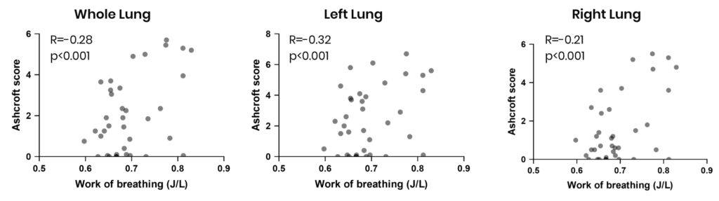
In the bleomycin model, normalized work of breathing increased following fibrosis induction. However, when work of breathing was compared with histological fibrosis scores, the relationship was weak. This is an important finding, as it suggests that histology can provide valuable information about the extent of fibrosis but is unable, by itself, to predict the amount of effort required to ventilate the lungs. | 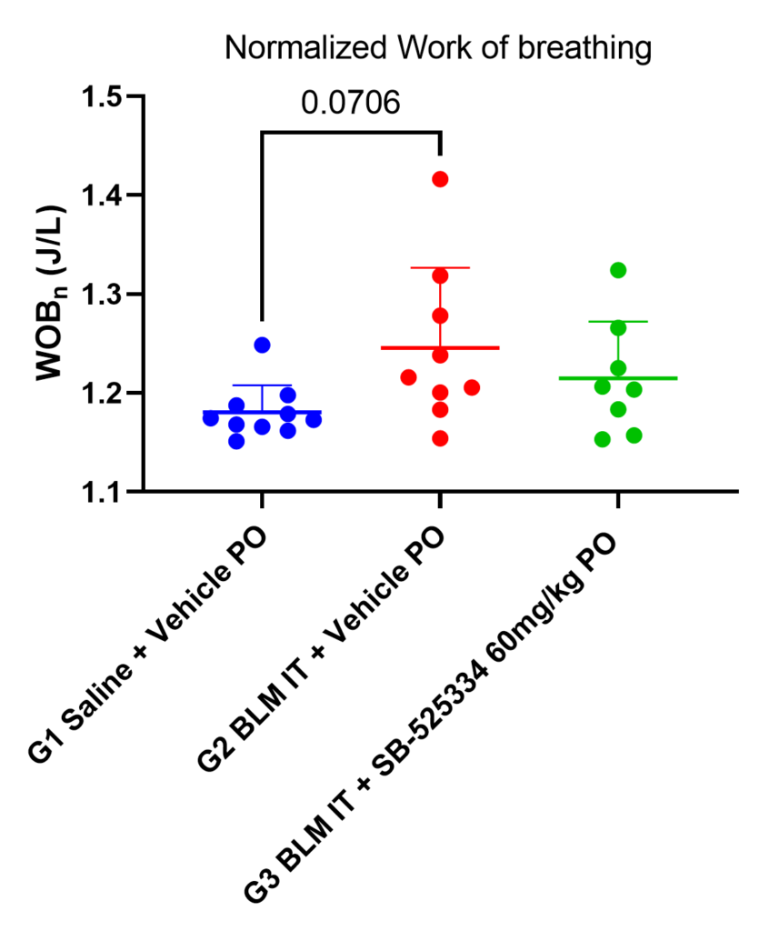 |
| Pressure-volume loops Lung function data can be visualized using pressure volume loops, which mimics the inspiration-expiration cycle. |
Here we can see that the untreated bleomycin group (red) has altered curves compared to the control group (blue), reaching a lesser volume at the end of a normal inspiration. The bleomycin group treated with SB-525334 (green) show a partial correction of this defect. | 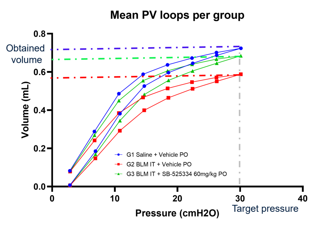 |
The objective of this study was to evaluate whether histology can stand on its own, or if introducing functional readouts in IPF studies is a necessary de-risking step before moving to clinical development. As such, we need to question whether histological parameters can reliably and strongly predict functional readout, and thus whether histological data alone can generate enough confidence in functional improvement to proceed to clinical trials with a candidate compound.
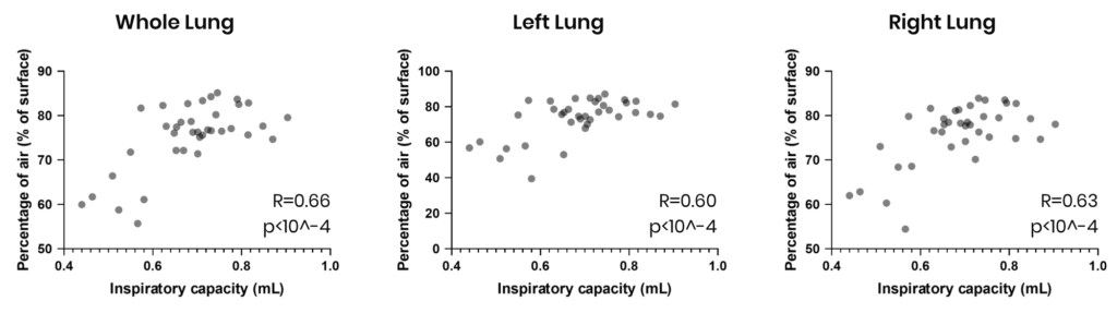
Can histology reliably predict inspiratory capacity? When comparing histological and functional results for inspiratory capacity, we can see a moderate correlation of +0.60. Correlation is impacted if both lungs are not evaluated using the same assessment:
Can histology reliably predict static compliance? In this study, Ashcroft scoring shows moderately strong negative correlations with functional static compliance, with reliability again decreasing if only one lung is assessed. Outlying datapoints reduce confidence that a change in the Ashcroft score is satisfactorily representative of a change in functional static compliance:
Can histology reliably predict normalized work of breathing? Correlations between Ashcroft scores and normalized work of breathing (or effort required to breathe) were very weak:
In summary, treatment with the ALK5 inhibitor resulted in a marked improvement in histological markers of fibrosis, including reduced collagen deposition and lower Ashcroft scores. However, improvements in lung function were considerably less pronounced, with only marginal changes observed in static compliance, inspiratory capacity, and work of breathing. Correlation analyses further demonstrated that histological endpoints were only moderate predictors of static compliance and inspiratory capacity and showed weak predictive value for work of breathing. These findings indicate that improvements in tissue pathology do not necessarily translate into proportional recovery of lung function, underscoring the importance of incorporating functional readouts alongside histological assessments in preclinical lung fibrosis studies.
Selecting endpoints for preclinical pulmonary fibrosis studies.
When selecting endpoints, researchers should always consider how their compound will ultimately be evaluated in patients, and which combination of preclinical endpoints will provide the clearest evidence that it will produce the intended therapeutic effect.
Histology remains a cornerstone of preclinical pulmonary fibrosis research, providing valuable insight into the extent and distribution of fibrotic pathology. However, improvements in tissue structure should not automatically be interpreted as equivalent improvements in lung function. This becomes particularly important when evaluating therapeutic efficacy. A compound may substantially reduce collagen deposition or improve fibrosis scores without producing an equivalent improvement across all aspects of respiratory function.
Conversely, depending on its mechanism of action, a therapeutic intervention could potentially improve a functional parameter without producing an equally substantial change in the biomarkers or histological features being measured.
Rather than viewing histological and functional assessments as competing measures of efficacy, they should be considered complementary components of a more complete evaluation. Histology demonstrates whether the underlying pathology has changed, while functional measurements help determine what those changes mean for the performance of the organ and, ultimately, their potential clinical relevance.
The question is therefore not whether histology or functional assessment is the better endpoint. We are better served by asking what each endpoint contributes to understanding therapeutic efficacy, and at what point the weight of evidence is sufficient to confidently move a candidate into clinical trials.
Key takeaways for researchers:
- Histological improvement does not necessarily predict functional recovery. Reductions in fibrosis and collagen deposition may not be matched by equivalent improvements across measures of lung function.
- Different functional endpoints answer different questions. Inspiratory capacity, static compliance and work of breathing capture distinct aspects of respiratory impairment and therapeutic response.
- Correlation should not be mistaken for prediction. Histological parameters may be associated with lung function without reliably predicting the magnitude of functional improvement.
- Select endpoints according to the translational question. Consider how therapeutic efficacy will ultimately be evaluated clinically and which aspects of that response can be meaningfully assessed preclinically.
- Combine complementary evidence where appropriate. Histology and functional assessment can together provide a more complete understanding of therapeutic activity than either approach alone.